CD44: A Multifunctional Mediator of Cancer Progression
Malak Hassn Mesrati, Saiful Effendi Syafruddin, M. Aiman Mohtar and 1 more
Biomolecules · 2021 · Review · CC BY
doi:10.3390/biom11121850 (opens in a new tab)
We email you when a new notice or a question about this paper appears.
Abstract
CD44, a non-kinase cell surface transmembrane glycoprotein, has been widely implicated as a cancer stem cell (CSC) marker in several cancers. Cells overexpressing CD44 possess several CSC traits, such as self-renewal and epithelial-mesenchymal transition (EMT) capability, as well as a resistance to chemo- and radiotherapy. The CD44 gene regularly undergoes alternative splicing, resulting in the standard (CD44s) and variant (CD44v) isoforms. The interaction of such isoforms with ligands, particularly hyaluronic acid (HA), osteopontin (OPN) and matrix metalloproteinases (MMPs), drive numerous cancer-associated signalling. However, there are contradictory results regarding whether high or low CD44 expression is associated with worsening clinicopathological features, such as a higher tumour histological grade, advanced tumour stage and poorer survival rates. Nonetheless, high CD44 expression significantly contributes to enhanced tumourigenic mechanisms, such as cell proliferation, metastasis, invasion, migration and stemness; hence, CD44 is an important clinical target. This review summarises current research regarding the different CD44 isoform structures and their roles and functions in supporting tumourigenesis and discusses CD44 expression regulation, CD44-signalling pathways and interactions involved in cancer development. The clinical significance and prognostic value of CD44 and the potential of CD44 as a therapeutic target in cancer are also addressed.
Figures7
From PubMed Central, U.S. National Library of Medicine.
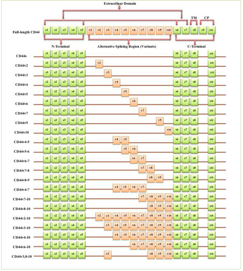
Figure 1
Full legend
Schematic diagram of the mouse CD44 gene and most CD44 isoforms involved in cancer progression. The full-length CD44 gene contains 20 exons in mice and 19 exons in humans, with the constant exons 1–5 and 16–20 encoding the N-terminal and the C-terminal domains. CD44 standard (CD44s) is encoded by these ten constant exons and contains no variant exons, whereas the CD44 variant (CD44v) is produced by the alternative splicing of a variable insertion of nine extra exons in humans or ten extra exons in mice. These extra exons are exons 6-15, typically identified as (v1 to v10) in mice and the exons 7-15 identified as (v2 to v10) in humans and are located between the N-terminal and C-terminal domains. CD44v can contain one or multiple variant exons and exon 19 is spliced out in all CD44 isoforms. Abbreviations: CD44s, CD44 standard; CD44v, CD44 variant; s, standard; v, variant; TM, transmembrane; CP, cytoplasmic. Green boxes refer to the constant/standard exons. Orange boxes refer to the variant exons.
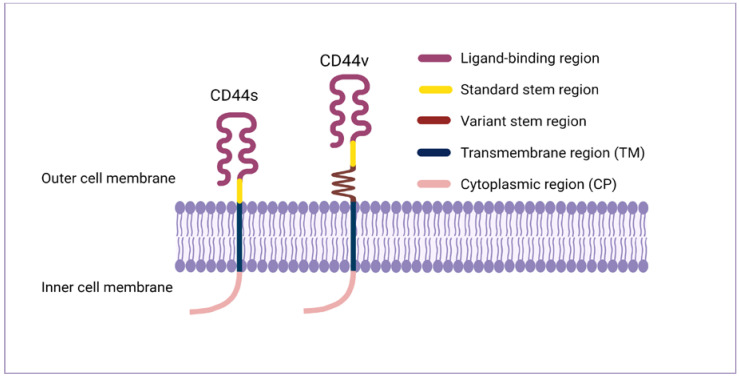
Figure 2
Full legend
CD44 protein structure. The CD44 protein has four primary regions: the extracellular region consists of seven extracellular domains including N-terminal domains (ligand-binding region), the stem region (variable stem region and/or standard stem region) which is the alternative splicing area containing an insertion of one or more variant exons, the transmembrane region (TM), and the C-terminal cytoplasmic (CP) region.
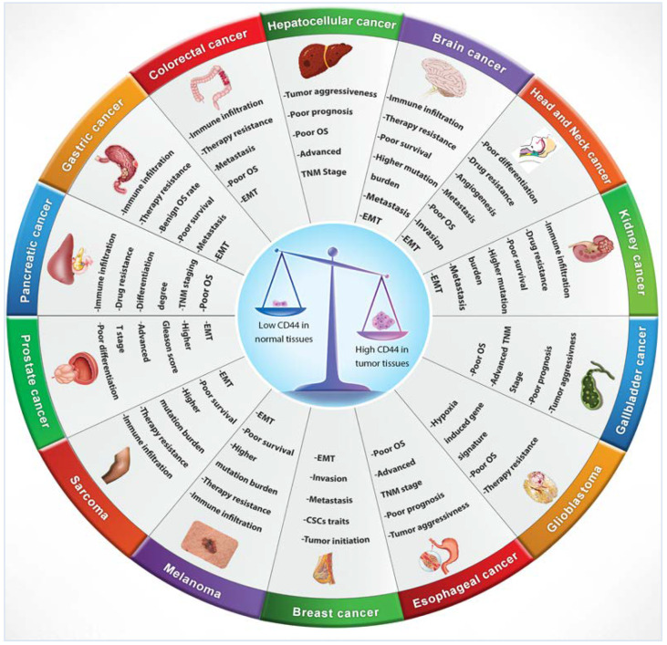
Figure 3
Full legend
CD44 distribution in normal versus cancerous tissues and its correlation with clinical outcomes.
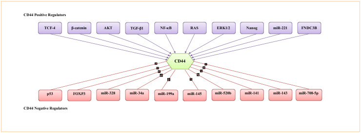
Figure 4
Full legend
Representative transcription factors, protein kinases, cytokines and miRNA involved in the regulation of CD44 activity.
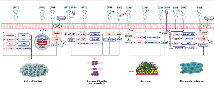
Figure 5
Full legend
Cancer-associated signalling pathways modulated by CD44. CD44 can upregulate EMT biomarkers, Snail1 and Zeb1, promoting proliferation and invasion. CD44 activates KRAS through the MAPK pathway, hence promoting tumour cell proliferation and survival. CD44 mediates invasion and metastasis by binding to HER2, leading to the inhibition of miR-139 and upregulation of CXCR4. There is positive crosstalk between CD44 and FGFR2 to maintain cancer stemness. CD44 regulates tumour cell proliferation, invasion and migration by modulating c-Src via AKT/GSK-3β signalling. CD44/VCAM-1 interaction promotes invasion signalling by the ezrin/PI3K pathway. CD44 binds to HA, resulting in stemness development via the PI3K/4EBP1/SOX2 pathway. Also, the HA/CD44 interaction can drive tumour invasion, metastasis and stemness through Src activating RAS/FAK/ERK pathways. Similarly, HA/CD44 stimulates the PI3K/AKT signalling pathway to increase therapeutic resistance. CD44 can sustain EGFR and AKT signalling by inhibition of Rab7A, leading to therapeutic resistance. CD44 promotes the expression of HAS2 by activating the PI3K/AKT signalling pathway. HAS2 further enhances CD44-mediated PI3K/AKT signalling, thus creating a positive feedback loop that drives tumour cell resistance and survival. CD44 can stabilise the cystine/glutamate antiporter (xCT), leading to increased GSH and decreased ROS levels, which, in turn, results in tumour cell proliferation and therapeutic resistance mediated by suppression of the p38 pathway. CD44 regulates tumour cell proliferation by the Wnt/β-catenin signalling pathway. CD44 can promote invasion and migration through the activation of the Hippo-YAP oncogene signalling pathway. CD44 mediates tumour cells resistance by upregulating Twist1 and AKT signalling. CD44-ICD binds to CREB, enhances S133 phosphorylation and enriches CREB recruitment to the cyclin D1 promoter, thus promoting cyclin D1 activity, resulting in cell proliferation. CD44-ICD is released in a hypoxic environment and binds to HIF-2α leading to induced stemness. Abbreviations: EMT, epithelial–mesenchymal transition; HER2, human epidermal growth factor receptor 2; CXCR4, C-X-C chemokine receptor type 4; FGFR2, fibroblast growth factor receptor 2; EGFR, epidermal growth factor receptor; HAS2, hyaluronan synthase 2; GSH, glutathione; ROS, reactive oxygen species; CD44-ICD, CD44 intracellular domain; CREB, cAMP response element-binding; HIF-2α, hypoxia-inducible factors 2 alpha.
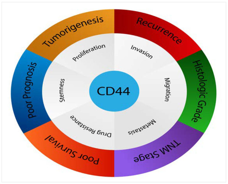
Figure 6
Full legend
CD44 affects numerous pathological processes in cancers.
Show all 7 figures
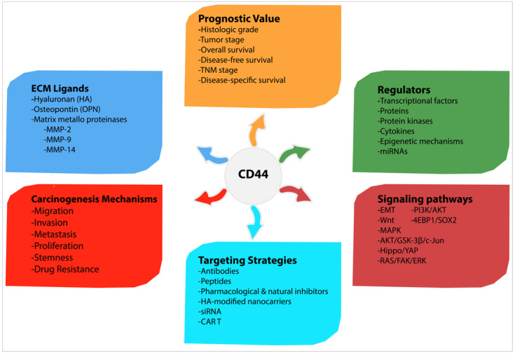
Figure 7
Full legend
Summary of carcinogenic mechanisms and signalling pathways induced by CD44, as well as CD44 regulators, ligands, prognostic value and possible targeting strategies.
References233
- 1.
- 2.
- 3.
Dalchau R., Kirkley J., Fabre J.W. Monoclonal antibody to a human brain-granulocyte-T lymphocyte antigen probably homologous to the W 3/13 antigen of the rat. Eur. J. Immunol. 1980;10:745-749.
Not in our library10.1002/eji.1830101004 (opens in a new tab)
- 4.
Hughes E.N., Mengod G., August J.T. Murine cell surface glycoproteins. Characterization of a major component of 80,000 daltons as a polymorphic differentiation antigen of mesenchymal cells. J. Biol. Chem. 1981;256:7023-7027.
Not in our library10.1016/s0021-9258(19)69093-9 (opens in a new tab)
- 5.
Stamenkovic I., Amiot M., Pesando J.M., Seed B. A lymphocyte molecule implicated in lymph node homing is a member of the cartilage link protein family. Cell. 1989;56:1057-1062.
Not in our library10.1016/0092-8674(89)90638-7 (opens in a new tab)
- 6.
Goodfellow P.N., Banting G., Wiles M.V., Tunnacliffe A., Parkar M., Solomon E., Dalchau R., Fabre J.W. The gene, MIC4, which controls expression of the antigen defined by monoclonal antibody F10.44.2, is on human chromosome 11. Eur. J. Immunol. 1982;12:659-663.
Not in our library10.1002/eji.1830120807 (opens in a new tab)
- 7.
Colombatti A., Hughes E.N., Taylor B.A., August J.T. Gene for a major cell surface glycoprotein of mouse macrophages and other phagocytic cells is on chromosome 2. Proc. Natl. Acad. Sci. USA. 1982;79:1926-1929.
Not in our library10.1073/pnas.79.6.1926 (opens in a new tab)
- 8.
Zeilstra J. CD44 Isoforms in Intestinal Cancer: Identity and Functions. Ph.D. Thesis. 2020.
Not in our library
- 9.
Ouhtit A., Rizeq B., Saleh H.A., Rahman M.M., Zayed H. Novel CD44-downstream signaling pathways mediating breast tumor invasion. Int. J. Biol. Sci. 2018;14:1782-1790.
Not in our library10.7150/ijbs.23586 (opens in a new tab)
- 10.
Loh T.J., Moon H., Cho S., Jang H., Liu Y.C., Tai H., Jung D.W., Williams D.R., Kim H.R., Shin M.G., et al. CD44 alternative splicing and hnRNP A1 expression are associated with the metastasis of breast cancer. Oncol. Rep. 2015;34:1231-1238.
Not in our library10.3892/or.2015.4110 (opens in a new tab)
- 11.
Rall C.J., Rustgi A.K. CD44 isoform expression in primary and metastatic pancreatic adenocarcinoma. Cancer Res. 1995;55:1831-1835.
Not in our library
- 12.
Mishra M.N., Chandavarkar V., Sharma R., Bhargava D. Structure, function and role of CD44 in neoplasia. J. Oral Maxillofac. Pathol. 2019;23:267-272.
Not in our library10.4103/jomfp.jomfp_246_18 (opens in a new tab)
- 13.
Tölg C., Hofmann M., Herrlich P., Ponta H. Splicing choice from ten variant exons establishes CD44 variability. Nucleic Acids Res. 1993;21:1225-1229.
Not in our library10.1093/nar/21.5.1225 (opens in a new tab)
- 14.
Screaton G.R., Bell M.V., Jackson D.G., Cornelis F.B., Gerth U., Bell J.I. Genomic structure of DNA encoding the lymphocyte homing receptor CD44 reveals at least 12 alternatively spliced exons. Proc. Natl. Acad. Sci. USA. 1992;89:12160-12164.
Not in our library10.1073/pnas.89.24.12160 (opens in a new tab)
- 15.
Zeilstra J., Joosten S.P., van Andel H., Tolg C., Berns A., Snoek M., Van De Wetering M., Spaargaren M., Clevers H., Pals S.T. Stem cell CD44v isoforms promote intestinal cancer formation in Apc(min) mice downstream of Wnt signaling. Oncogene. 2014;33:665-670.
Not in our library10.1038/onc.2012.611 (opens in a new tab)
- 16.
Goodison S., Urquidi V., Tarin D. CD44 cell adhesion molecules. Mol. Pathol. 1999;52:189-196.
Not in our library10.1136/mp.52.4.189 (opens in a new tab)
- 17.
Chen K.L., Li D., Lu T.X., Chang S.W. Structural Characterization of the CD44 Stem Region for Standard and Cancer-Associated Isoforms. Int. J. Mol. Sci. 2020;21:336.
Not in our library10.3390/ijms21010336 (opens in a new tab)
- 18.
- 19.
Liu X., Taftaf R., Kawaguchi M., Chang Y.F., Chen W., Entenberg D., Zhang Y., Gerratana L., Huang S., Patel D.B., et al. Homophilic CD44 Interactions Mediate Tumor Cell Aggregation and Polyclonal Metastasis in Patient-Derived Breast Cancer Models. Cancer Discov. 2019;9:96-113.
Not in our library10.1158/2159-8290.cd-18-0065 (opens in a new tab)
- 20.
Wolf K.J., Shukla P., Springer K., Lee S., Coombes J.D., Choy C.J., Kenny S.J., Xu K., Kumar S. A mode of cell adhesion and migration facilitated by CD44-dependent microtentacles. Proc. Natl. Acad. Sci. USA. 2020;117:11432-11443.
Not in our library10.1073/pnas.1914294117 (opens in a new tab)
- 21.
Hassn Mesrati M., Behrooz A.B., Abuhamad A.Y., Syahir A. Understanding Glioblastoma Biomarkers: Knocking a Mountain with a Hammer. Cells. 2020;9:1236.
Not in our library10.3390/cells9051236 (opens in a new tab)
- 22.
Ludwig N., Szczepanski M.J., Gluszko A., Szafarowski T., Azambuja J.H., Dolg L., Gellrich N.-C., Kampmann A., Whiteside T.L., Zimmerer R.M. CD44(+) tumor cells promote early angiogenesis in head and neck squamous cell carcinoma. Cancer Lett. 2019;467:85-95.
Not in our library10.1016/j.canlet.2019.10.010 (opens in a new tab)
- 23.
Gzil A., Zarębska I., Bursiewicz W., Antosik P., Grzanka D., Szylberg Ł. Markers of pancreatic cancer stem cells and their clinical and therapeutic implications. Mol. Biol. Rep. 2019;46:6629-6645.
Not in our library10.1007/s11033-019-05058-1 (opens in a new tab)
- 24.
Durko L., Wlodarski W., Stasikowska-Kanicka O., Wagrowska-Danilewicz M., Danilewicz M., Hogendorf P., Strzelczyk J., Malecka-Panas E. Expression and Clinical Significance of Cancer Stem Cell Markers CD24, CD44, and CD133 in Pancreatic Ductal Adenocarcinoma and Chronic Pancreatitis. Dis. Markers. 2017;2017:3276806.
Not in our library10.1155/2017/3276806 (opens in a new tab)
- 25.
Li W., Qian L., Lin J., Huang G., Hao N., Wei X., Wang W., Liang J. CD44 regulates prostate cancer proliferation, invasion and migration via PDK1 and PFKFB4. Oncotarget. 2017;8:65143-65151.
Not in our library10.18632/oncotarget.17821 (opens in a new tab)
- 26.
Wang Z., Tang Y., Xie L., Huang A., Xue C., Gu Z., Wang K., Zong S. The Prognostic and Clinical Value of CD44 in Colorectal Cancer: A Meta-Analysis. Front. Oncol. 2019;9:309.
Not in our library10.3389/fonc.2019.00309 (opens in a new tab)
- 27.
Holah N.S., Aiad H.A., Asaad N.Y., Elkhouly E.A., Lasheen A.G. Evaluation of the role of CD44 as a cancer stem cell marker in colorectal carcinoma: Immunohistochemical study. Menoufia Med. J. 2017;30:174-183.
Not in our library10.7860/jcdr/2017/22671.9291 (opens in a new tab)
- 28.
Zhang H., Brown R.L., Wei Y., Zhao P., Liu S., Liu X., Deng Y., Hu X., Zhang J., Gao X.D., et al. CD44 splice isoform switching determines breast cancer stem cell state. Genes Dev. 2019;33:166-179.
Not in our library10.1101/gad.319889.118 (opens in a new tab)
- 29.
Zhao P., Xu Y., Wei Y., Qiu Q., Chew T.L., Kang Y., Cheng C. The CD44s splice isoform is a central mediator for invadopodia activity. J. Cell Sci. 2016;129:1355-1365.
Not in our library10.1242/jcs.171959 (opens in a new tab)
- 30.
Brown R.L., Reinke L.M., Damerow M.S., Perez D., Chodosh L.A., Yang J., Cheng C. CD44 splice isoform switching in human and mouse epithelium is essential for epithelial-mesenchymal transition and breast cancer progression. J. Clin. Investig. 2011;121:1064-1074.
Not in our library10.1172/jci44540 (opens in a new tab)
- 31.
Mima K., Okabe H., Ishimoto T., Hayashi H., Nakagawa S., Kuroki H., Watanabe M., Beppu T., Tamada M., Nagano O., et al. CD44s regulates the TGF-β-mediated mesenchymal phenotype and is associated with poor prognosis in patients with hepatocellular carcinoma. Cancer Res. 2012;72:3414-3423.
Not in our library10.1158/0008-5472.can-12-0299 (opens in a new tab)
- 32.
Miwa T., Nagata T., Kojima H., Sekine S., Okumura T. Isoform switch of CD44 induces different chemotactic and tumorigenic ability in gallbladder cancer. Int. J. Oncol. 2017;51:771-780.
Not in our library10.3892/ijo.2017.4063 (opens in a new tab)
- 33.
Chen Q., Gu M., Cai Z.k., Zhao H., Sun S.C., Liu C., Zhan M., Chen Y.B., Wang Z. TGF-β1 promotes epithelial-to-mesenchymal transition and stemness of prostate cancer cells by inducing PCBP1 degradation and alternative splicing of CD44. Cell. Mol. Life Sci. 2021;78:949-962.
Not in our library10.1007/s00018-020-03544-5 (opens in a new tab)
- 34.
Zhao S., Chen C., Chang K., Karnad A., Jagirdar J., Kumar A.P., Freeman J.W. CD44 Expression Level and Isoform Contributes to Pancreatic Cancer Cell Plasticity, Invasiveness, and Response to Therapy. Clin. Cancer Res. 2016;22:5592-5604.
Not in our library10.1158/1078-0432.ccr-15-3115 (opens in a new tab)
- 35.
Tsubouchi K., Minami K., Hayashi N., Yokoyama Y., Mori S., Yamamoto H., Koizumi M. The CD44 standard isoform contributes to radioresistance of pancreatic cancer cells. J. Radiat. Res. 2017;58:816-826.
Not in our library10.1093/jrr/rrx033 (opens in a new tab)
- 36.
Ozawa M., Ichikawa Y., Zheng Y.W., Oshima T., Miyata H., Nakazawa K., Guan H.B., Shiozawa M., Akaike M., Watanabe K., et al. Prognostic significance of CD44 variant 2 upregulation in colorectal cancer. Br. J. Cancer. 2014;111:365-374.
Not in our library10.1038/bjc.2014.253 (opens in a new tab)
- 37.
Gotoda T., Matsumura Y., Kondo H., Saitoh D., Shimada Y., Kosuge T., Kanai Y., Kakizoe T. Expression of CD44 variants and its association with survival in pancreatic cancer. Jpn. J. Cancer Res. 1998;89:1033-1040.
Not in our library10.1111/j.1349-7006.1998.tb00493.x (opens in a new tab)
- 38.
Todoroki K., Ogasawara S., Akiba J., Nakayama M., Naito Y., Seki N., Kusukawa J., Yano H. CD44v3+/CD24- cells possess cancer stem cell-like properties in human oral squamous cell carcinoma. Int. J. Oncol. 2016;48:99-109.
Not in our library10.3892/ijo.2015.3261 (opens in a new tab)
- 39.
Holm F., Hellqvist E., Mason C.N., Ali S.A., Delos-Santos N., Barrett C.L., Chun H.J., Minden M.D., Moore R.A., Marra M.A., et al. Reversion to an embryonic alternative splicing program enhances leukemia stem cell self-renewal. Proc. Natl. Acad. Sci. USA. 2015;112:15444-15449.
Not in our library10.1073/pnas.1506943112 (opens in a new tab)
- 40.
Bánky B., Rásó-Barnett L., Barbai T., Tímár J., Becságh P., Rásó E. Characteristics of CD44 alternative splice pattern in the course of human colorectal adenocarcinoma progression. Mol. Cancer. 2012;11:83.
Not in our library10.1186/1476-4598-11-83 (opens in a new tab)
Cited by papers we hold4
Reads
Read by people on this page
None yet.
Checked or read by AI through naulon
None yet.
A person counts once a day per paper. We keep no cookie and no address: a reader is a hash that changes every day. Any month under five reads shows no number.
Are you an author?
Is this your paper?
Claim it to add your note and be paid when agents cite it. Free.