The molecular mechanism of nuclear transport revealed by atomic-scale measurements
Loren E Hough, Kaushik Dutta, Samuel Sparks and 5 more
eLife · 2015 · CC BY
doi:10.7554/elife.10027 (opens in a new tab)
We email you when a new notice or a question about this paper appears.
Abstract
Nuclear pore complexes (NPCs) form a selective filter that allows the rapid passage of transport factors (TFs) and their cargoes across the nuclear envelope, while blocking the passage of other macromolecules. Intrinsically disordered proteins (IDPs) containing phenylalanyl-glycyl (FG)-rich repeats line the pore and interact with TFs. However, the reason that transport can be both fast and specific remains undetermined, through lack of atomic-scale information on the behavior of FGs and their interaction with TFs. We used nuclear magnetic resonance spectroscopy to address these issues. We show that FG repeats are highly dynamic IDPs, stabilized by the cellular environment. Fast transport of TFs is supported because the rapid motion of FG motifs allows them to exchange on and off TFs extremely quickly through transient interactions. Because TFs uniquely carry multiple pockets for FG repeats, only they can form the many frequent interactions needed for specific passage between FG repeats to cross the NPC.
Full text
From PubMed Central, U.S. National Library of Medicine, as current on Oct 24, 2026. Paragraph numbers are ours, so an answer can point at one.
Introduction
Nuclear pore complexes (NPCs) are the sole mediators of bi-directional nucleocytoplasmic trafficking. Transport is rapid and reversible, with the entire process of transport factor (TF) docking, passage and release across the NPC taking only a few milliseconds. The NPC consists of a ∼30-nm diameter central channel filled with phenylalanyl-glycyl-repeat-rich nucleoporins (FG Nups) which provide the selective filter. Depletion or deletion of the FG Nups results in leaky, non-selective barriers. Moreover, the FG domains in isolation facilitate selective passage of TFs through nanopores or accumulation in hydrogels.
It is generally accepted that TF interaction with FG repeats reduce the diffusional barrier to enable selective transport, though the molecular mechanism of TF passage through the NPC remains largely unknown. TFs may alter the properties of the FG permeability barrier. Mesoscale observations of TF-FG repeat interactions in vitro have shown that TFs can change the height of FG brushes on planar surfaces, modulate the transport of inert cargo, assemble with FG Nups into large assemblies, and inhibit amyloid hydrogel formation observed for some FG Nups. Whether these behaviors arise from changes in FG structure or as a result of the multivalent FG-TF interaction remains undetermined, as do their contributions to in vivo nuclear transport.
Several mesoscale models have attempted to explain how FG Nups prevent the passage of most macromolecules while allowing selective transport of TFs alone and with their cargo. The FG Nups have been proposed to form a selective barrier due to their reversible inter-chain cohesion (hydrogel and bundle models), entropic exclusion (virtual gating model), collapse upon TF binding (reduction in dimensionality model), or a combination thereof (forest model). These models differ in their predictions of FG behavior on their own (ranging from highly mobile to fully self-associated) and upon binding of TFs. For example, the hydrogel, forest and reduction in dimensionality models invoke large changes in FG Nup behavior upon TF interaction. Crucially, none of these models or current observations fully explains how transport can be both selective and rapid, as is seen in vivo.
These conflicting models of the mechanism of NPC selectivity remain because of a lack of atomic-scale experimental data describing FG Nup behaviors and interactions. Therefore, we used nuclear magnetic resonance (NMR) techniques to provide a rich readout of FG Nup behaviors at atomic detail (Figure 1A). We defined a minimal system including FG attachment, FG repeat type, and TF interaction, the essential features necessary to recapitulate selective transport in vitro. We measured the physical state of FG repeats with and without TFs bound. Because environment strongly affects intrinsically disordered proteins (IDPs), including FG Nups, we mimicked the normal environment of NPCs using Xenopus egg extract, the best characterized environment for in vitro nuclear transport measurements. In addition, we tested the following: cytoplasm of living Escherichia coli using in cell NMR; E. coli high speed lysate; and buffer alone, the latter lacking crowding agents or competitors and being the milieu in which these proteins have been most studied previously.
We studied Nsp1, the most tested and characterized FG Nup which has been shown in vitro to mimic transport faithfully. We focused on two segments of Nsp1 for which there is a consensus that they prototypically represent the extreme flavors and behaviors of FG Nups; (i) the N-terminal segment of low charge (Asn-rich) and irregularly spaced FG repeats observed to be highly cohesive and form amyloid hydrogels under certain conditions (FG-N) and (ii) the central segment of significant charge (Lys-rich) with regular FSFG repeats typically observed to be highly soluble (FSFG-K). The isolated N-terminal domain forms selective hydrogels in vitro and is able to replace Nup98-FG domain in reconstituted Xenopus nuclei to provide the primary barrier enabling selective transport. We studied these fragments individually and in combination in a wide range of constructs and conditions, including where the FG repeats are tethered to mimic their arrangement in the NPC (Table 1, Figure 1—figure supplement 1).
The cellular milieu maintains FG Nups as highly dynamic IDPs
FG Nups were fully disordered and highly dynamic in all cellular milieu tested. In NMR measurements, the degree of secondary structure is correlated with the difference between the 13C chemical shift values of the α and β carbons relative to random coil values for each residue. The degree of secondary structure quantified in this way is near zero for all residues in FG-N and FSFG-K constructs in all conditions tested (Figure 1B, Figure 1—figure supplement 1). This behavior of intrinsic disorder, judged from chemical shifts, is strikingly robust, being seen under a wide variety of conditions and also (for FSFG-K constructs) in buffers of varying pH (Figure 1—figure supplement 3).
Our observations in cellular milieux are in contrast to the behavior of the constructs containing the FG-N domain in buffer alone. Under these conditions, dynamic light scattering and NMR measurements indicate that FG-N forms a hydrogel-like material (Figure 1—figure supplement 4). The in buffer behavior of our construct is consistent with extensive previous observations of β-sheet formation and aggregation of the N-terminal domain of Nsp1 (FG-N). However, when FG-N constructs are observed by NMR inside a living cell, or in the presence of cell lysates or mimics, there is no appearance of high molecular weight components—NMR spectra of FG-N constructs in living E. coli did not change with time over more than 24 hr (Figure 1—figure supplement 5). These data indicate that the cellular milieu is a strong inhibitor of intermolecular FG repeat aggregation. The state of FG repeats in our system is therefore highly dynamic, as has been reported in vivo.
To understand the differences between protein structure and dynamics in cellular mimics as compared to buffer, we measured spin relaxation parameters (R1, R2, nOe) which quantify the motion and interactions of the residues on timescales of μs to ms (R1, R2,) and ps to ns (nOe). NMR relaxation properties of FG Nups are significantly different between buffer alone and a protein-rich environment (Figure 1C). We measured large increases in R2/R1, indicative of transient spectral changes on interaction between the constructs and the cellular milieu (Figure 1C, Figure 1—figure supplement 2). The increases in R2/R1 were not seen upon increasing viscosity or crowding by inert agents (Figure 1—figure supplement 6), indicating that these changes result from weak binding of the FG repeats to the proteinaceous milieu as suggested by previous studies. The phenylalanines and their adjacent residues show the primary interactions, with the greatest increases in R2/R1, while the spacer sequences remained relatively unaffected. The nuclear Overhauser effect (nOe) data show that the FG-N are highly mobile and flexible (Figure 1C, Figure 1—figure supplement 2), indicating no sequence-specific compaction, folding, or molten globule formation. The cellular milieu is thus in a state of constant, non-specific, dynamic interaction with the FG repeats.
Our results suggest that interactions of FG repeats with the cellular milieu stabilize the unfolded state by engaging the hydrophobic phenylalanines in transient interactions that decrease their contact with the water, reducing the driving force for hydrophobic collapse and amyloid formation. This is consistent with previous work that demonstrated that mutation of the Fs to more hydrophilic residues inhibits aggregation. Thus, exchange with the cellular milieu maintains the FG Nups in a highly dynamic, disordered state by inhibiting the intramolecular interactions that lead to aggregation in buffer.
Random fuzzy interactions with transport factors
We used the atomic-level readout of our system to interrogate key aspects of FG Nup-TF interactions that have previously been inaccessible. It has long been established using crystallographic, computational, and NMR approaches that FG residues bind to hydrophobic pockets on TFs (reviewed in ). However, the dynamics of this interaction and the behavior of the spacer regions between FG residues upon TF binding have remained uninterrogated, leading to proposals ranging from remaining disordered to significant structural rearrangements. Our assays provide a direct readout of the dynamics of the interaction and the state of the linker regions upon TF binding (Figure 2).
Many previous in vitro observations of FG-TF interactions lacked demonstration of reversibility and showed strong affinities, incompatible with the rapid transport rates observed in vivo. By monitoring the recovery of the NMR spectral characteristics on dilution of Kap95, the interacting TF, we showed that the FG repeat-TF interactions are fully reversible in our system (Figure 2—figure supplement 1).
We used measured changes in amide resonances peak position and intensity as a function of Kap95 concentration using 2D Heteronuclear Single Quantum Coherence (HSQC) experiments (Figure 2A). Each (non-proline) residue contributes a peak, with the amide proton resonance frequency on the x-axis and nitrogen resonance frequency on the y-axis. The signal intensity of each peak is sensitive to the mobility and environment of that residue; upon binding to a TF, the effects of slower motion and of differing environment result in the signal of the corresponding peak decreasing. Strikingly, although the phenylalanine repeats themselves are attenuated upon Kap95 interaction, the majority of peaks are minimally affected, showing that the corresponding residues remain disordered and dynamic (Figure 2A). The spacer regions are thus highly mobile even within the interacting state. An extreme of this observation is seen for K332, which shows no attenuation (left subpanel in Figure 2A, lowest trace Figure 2C), and thus this residue remains flexible both in free and when bound to TF. When we examined attenuation as a function of residue number, we observed a striking periodic pattern of minimal attenuation for most residues and significant attenuation for FG repeats (Figure 2B). The degree of signal attenuation falls off relatively uniformly at both sides of each FG repeat, consistent with the transient binding of individual FG residues, with the remaining residues remaining fully disordered and highly dynamic. The minimally attenuated residues include those that have previously been implicated in forming phase changes of the FG repeat regions. We did not observe any major state change of spacers resulting from TF addition, such as caused by the formation or breakage of secondary structure, or gel/sol transitions.
We observe that the binding interface between FG Nups and TFs at each interacting site is small—just the 2–4 residues surrounding the FG repeat itself—and the residues that do not interact directly with the TF sites remain highly mobile and dynamic. The size of the binding interface is consistent with other known transient interactions. We estimated a minimum effective affinity of Kap95 for the FSFG-K from a titration observing 15N R2 (Figure 2—figure supplement 2). The true Kd is greater than 36 μM, fully consistent with rapid, reversible transport.
Interaction or binding while remaining primarily disordered is highly unusual, as many functional IDPs are believed to adopt significant secondary structure formation upon interaction, which we do not see here. This behavior is reminiscent of that proposed for random fuzzy complexes, in which one partner remains dynamically disordered and transient interactions predominate during selective recognition.
FG repeat behavior is largely independent of packing
Many observations suggest that there are multiple transport routes across the NPC, with FG Nups arranged in a variety of geometries. Thus, because the NPC contains a large number of FG Nups in different arrangements to each other, we investigated the common features of the effects of local packing and attachment on FG repeats (Figure 3, Table 1). We designed chimeric proteins containing the FG domains in homotypic and heterotypic combinations. Some constructs included the tetramerization domain of p53, allowing us to form complexes that mimic the attachment of the FG domains to the NPC. We found essentially no change in chemical shifts and only modest changes in linewidth upon attachment to the p53 tetramerization domain (Figure 3). Any ordered protein of this size (∼120 kDa) would be invisible using our NMR experiments, demonstrating that FG Nups remain dynamic IDPs even in large complexes, as previously measured for other large IDPs. Notably, our largest constructs have a diameter measured by dynamic light scattering of 13 nm (Figure 3B), a significant fraction of the 30-nm diameter of the NPC. Our results reveal that the behavior of a given FG repeat is not affected strongly by the number or type of other FG repeats surrounding it (i.e., no ‘emergent properties’). Our results also strongly indicate that there are no significant FG–FG interactions, as interactions between different types of FGs should impact their average environment. However, we cannot completely exclude that extremely weak, dynamic interactions can occur between FG repeats, and indeed such interactions may modulate the long distance distribution of FG repeat regions in the NPC. The FG Nups thus remain a highly dynamic fluid even at sizes, arrangements, and packing densities commensurate with the NPC. We observed the same pattern of rapid exchange of the FG residues with TF binding sites while the spacer regions remain highly mobile in all environments studied and with all constructs tested, showing the robustness of our findings (Figure 4).
While all overall behaviors are similar between FG-N and FSFG-K segments, in monomers or in tetramers, there are some intriguing differences. For example, in constructs where both FG types are present (Figure 4), not all FG-N phenylalanine residues are completely attenuated. This may be evidence for specificity of certain FG repeat types to binding particular TFs.
FG Nups remain dynamic and disordered in their functional states
The FG repeats fill the central channel of the NPC to form a barrier to non-specific macromolecular diffusion. We show that, even when densely packed and in mixed flavors as in the NPC, the FG repeat regions studied here remain fully disordered and highly dynamic. The two domains studied represent the extreme behaviors observed for FG Nups in vitro: FSFG-K is highly soluble and non-cohesive, while FG-N in buffer is highly cohesive, a prototypical hydrogel-forming FG Nup. It is clearly possible to make FG repeats aggregate (Figure 1—figure supplements 4, 5), as has been demonstrated for many proteins. However, our data are inconsistent with any such hydrogel state in cellular milieu. Re-arrangements of internal non-covalent crosslinks within hydrogels are on slow (second to minute) timescales, long enough to form a solid gel resistant to deformation, and too slow for solution NMR analysis. Instead of such a highly internally interacting, slowly moving gel, our results demonstrate that FG repeat regions form a highly dynamic phase, consistent with the rapid rates of nuclear transport. A highly dynamic, fluid state for FG repeats in vivo is in agreement with measurements of the living NPC. FG Nups do not appear to form a molten globule or collapsed state, as has also been proposed, instead behaving as a fully disordered IDP. Our results also allow us to distinguish their IDP class. FG repeats do not appear to populate more folded states either inter- or intra-molecularly upon interaction with either TFs or other FG Nups. Instead, FG repeats lack any secondary structure, even when interacting with cognate binding partners, indicating that IDP mobility may be a key ingredient in nuclear transport.
Non-TF interactions of FG repeats
Our results highlight the importance of the cellular environment for determining the behavior of the FG Nups because multiple weak interactions with cellular components stabilize the FG Nups in a disordered state. We propose that the selectivity of the NPC is thus maintained by both non-specific and specific cellular interactions, consistent with recent work demonstrating that FG permeability barrier is modulated by TFs. We show that the modulation of permeability of FG brush behavior by TFs is not a result of structural changes of the FG Nups, but must result instead from the rapid, multivalent interactions of TFs with FG repeats.
Our data, strongly supporting that FG repeats are highly dynamic tethered IDPs, imply that they must form entropic bristles (i.e., strongly sterically hindered regions) around their tether site (the central channel of the NPC). This hindrance increases with increasing size of the passing macromolecules; hence, small proteins can pass more easily than large ones. These larger nonspecific macromolecules interact with the FG repeats with insufficient frequency to overcome the entropic barrier of the FG repeats' polymer bristle structure, thus being effectively excluded. TFs, however, have sufficient interaction frequency to do so—the original tenet of the ‘virtual gating’ idea, now delineated at the atomic scale by these results. In summary, the FG repeat regions, crowded around and within the central channel, may set up an entropic barrier that excludes macromolecules from their vicinity while permitting the approach of small molecules; however, macromolecules that interact with the FG repeats (such as TFs) can overcome this barrier by their multiple and frequent interactions with FG Nups. We are pursuing experiments which we hope in the near future will gage the precise nature and magnitude of the exclusion mechanism.
Extremely fast on and off rates provide an explanation for rapid transport
Our results suggest that FG disorder is critical to the speed of selective nucleocytoplasmic transport. Selectivity is determined by the free energy gain upon TF-NPC binding, with the off rate (and so speed of transport) constrained by koff = Kd·kon. For a given selectivity (Kd), the residence time within the NPC is constrained by the off rate. IDPs are able to engage far more rapidly than most ordered proteins, allowing for high on and off rates while maintaining selectivity. For FGs in particular, several key lines of evidence indicate that the interaction of FG Nups with TFs is extraordinarily rapid, that is, each FG motif is in extremely fast exchange with TFs. We have directly shown that the binding interface is only 2–4 amino acids (the FG repeat itself). The buried surface area of this interaction is <1000 Å2, consistent with other known transient interactions. The spacer regions remain highly mobile and the degree of signal attenuation falls off relatively uniformly at both sides of each FG repeat (Figure 2B), fully consistent with the transient binding of individual FG residues. Within the NPC, a TF is able to rapidly diffuse, engaging transiently with multiple FG motifs across different FG Nups. The association rate of TFs with individual FG motifs is very fast because the FGs move very rapidly and the TF has multiple binding pockets available for interaction. Taken together, these results indicate that the dynamics of FG-TF interaction are extraordinarily rapid. As a result, the interaction can be strong enough to be selective, and yet remain extremely fast, allowing rapid transit through the NPC.
In essence, it is the quantity, rather than quality, of interactions possible per molecule with FG repeats that distinguishes a TF from other macromolecules (Figure 5). Transient non-specific interactions of the FG repeats with their milieu help maintain the disordered and dynamic nature of the barrier. Given the low sequence and structural complexity of the FG repeats, such non-specific interactions are to be expected but are not strong enough for these non-specific macromolecules to overcome the FG Nups' diffusion barrier. In contrast, TFs bind specifically to the FG motifs through pockets tuned for this purpose while leaving the spacers between the motifs highly mobile. The fact that the spacers—and the FG repeats as a whole—remain fully mobile even when interacting with a TF also means that the barrier to non-specific passage formed by a dense fluid of FG repeats in the NPC remains fully intact, regardless of even large TF fluxes (a significant issue with models that invoke state changes, which our results and model circumvent).
While the FG repeats show little evidence of changed behavior upon oligomerization or TF interaction, it is likely that their structured attachment sites and diversity of flavors produce modulation of composition within the NPC. These variations are potentially important for the organization of alternate transport pathways. Our approach and its future extensions will thus be a powerful tool for the detailed characterization of these different nuclear transport pathways. The NPC is a biological example of the idiom ‘many hands make light work’; the combined effect of many weak, transient interactions provides a specific multifunctional transport system, which allows for the simultaneous passage of hundreds of different macromolecules with a wide range of sizes and for robustness to significant alterations in the cell.
Materials and methods
Proteins (Table 1) were expressed in BL21DE3 Gold cells using expression plasmids pET24a or pRSFDuet.
Protein concentration was determined by BCA analysis (Pierce Co.), except for Kap95 determined by OD280 based on standard amino acid content. IPTG was from Gold Biotechnology. Bovine Serum Albumin (BSA), Pepstatin, PMSF, Protease Inhibitor Cocktail are from Sigma. Xenopus egg extract was the generous gift of Takashi Onikubo and David Shechter. Lyophilized coli extract was prepared by modification of the preciously described method using E. coli BL21 Gold cells grown in LB to OD600 of 0.8, resuspended in Buffer A + 18 mg/l PMSF +0.4 mg/l pepstatin A, lysed by passing through a Microfluidizer, and centrifuged at 112,000 g for 30’ at 4°C.
Expression constructs and protein production
The FG-N, and FSFG-K segments were derived as previously described with the following modifications. Expression plasmids (pET24a, pRSFDuet) containing FG Nup fragments were transformed into BL21DE3 Gold cells (Agilent). Cells containing pET24a or pRSF constructs were grown to OD600 of 0.8, induced with 1 mM IPTG and harvested after 2–4 hr. The cells' periplasm was removed by osmotic shock, and lysed under denaturing conditions (8 M urea). Urea was maintained throughout the purification for FG-N, whereas FSFG-K had 8 M urea in the lysis, 3 M urea in the first wash, and no urea in the remaining washes and elution. Proteins were purified on Talon resin in 20 mM HEPES, 150 mM KCl, 2 mM MgCl2, (Buffer A, at pH 7.0), with protease inhibitor cocktail (PIC), PMSF, and pepstatin. Kap95 was prepared as previously described [U-15N] and [U-13C, 15N] materials were prepared using M9 media containing 15NH4Cl and [U-13C] glucose (Cambridge Isotopes Limited, MA) as needed. The tetramerization domain of p53 was used to provide intramolecular crowding by construction of fusions with the tetramerization segment. As with all our constructs, the preparation was analyzed by size exclusion chromatography (SEC) to ensure that we were examining un-aggregated protein of the correct size and quantity (data not shown) and confirmed by dynamic light scattering.
NMR measurements
Experiments were conducted on Bruker spectrometers at 700 MHz and 298 K unless otherwise indicated. In cell NMR methods used the general procedures developed for STINT-NMR, and other IDP studies. Assignment of resonances used the standard triple resonance approach as in;;;;, including HNCO, HNCACO, HNCACM, CBCACINH, HNCA, HNCOCA, CCCONH, and HCCONH. Assignments were conducted in cell (E. coli)/buffer E and in buffer A, for the sets tabulated in Table 2, and were confirmed in other buffers by examination of HSQCs. Relaxation methods used standard procedures. No corrections were applied for possible changes of fast exchange with solvent water, as described for R2(CPMG) for the following reasons. The observed changes for the IDP α-synuclein are less than two fold, and sequence-independent for a change of pH from 7.4 to 6.2 (Figure 1; ) while for FG Nups we observe up to c. sixfold sequence dependent variation of R2. Secondly, the same sequence dependent variations of R2 are seen in buffered E. coli extracts, with controlled pH (Figure 1—figure supplement 6). ‘Titration’ experiments were conducted at 800 or 700 MHz in buffers indicated, by preparation of separate samples for each change of concentration of Kap95.
Titration experiments for 15N R2 were conducted at 500 MHz in buffer A, by preparing separate samples. Kap95 was fixed at 20 μM and 15N FSFG-K concentration was varied from 12.5 μM to 1200 μM. Each sample was prepared by addition of standard volumes of stocks to obtain the concentrations. No additional normalization was used. Each R2 data acquisition used 6 delay points. For FSFG:Kap95 molar ratio of 1:1 and 0.625:1 the following delay times were used 0, 32.64, 65.28, 97.92, 130.56, 163.2 ms. For FSFG:Kap95 molar ratio of 4:1 the following delay times were used 0, 81.6, 163.2, 244.8, 326.4, 408 ms. For FSFG:Kap95 molar ratio of 7:1 the following delay times were used 0, 81.6, 163.2, 244.8, 326.4, 489.6 ms. For FSFG:Kap95 molar ratios of 60:1, 28.75:1 and 11:1 the following delay times were used 0, 81.6, 163.2, 326.4, 489.6, 652.8 ms. The same delays were used for an FSFG-N alone and the R2 values obtained provided the value of R2 free (R2(0)) in the binding equation. The numbers of scans were optimized for S/N for each sample and total acquisition time per sample was ∼<13 hr.
NMR analysis
The chemical shifts and their standard deviations were used in the standard equation to derive deviations from predicted shift values for the sequences of the assigned proteins. The values for these for Δδ13Cα- Δδ 13Cβ are shown in Figure 1—figure supplement 1.
For NMR relaxation analysis, we derive approximate correlation times for ps/ns, associated with the different correlation times for hetero nuclear nOe by a direct calculation method that, in part, normalizes across different observation fields/frequencies, avoiding any complexities of spectral density analysis (for example, ), of the unverified applicability of a ‘Model Free’ approach, and of analysis of simulated molecular dynamics which may not be generally applicable to all IDPs.
The apparent correlation times for motion of backbone amides was derived from use of the heteronuclear 15N[1H] nOe using the standard dipolar treatment, ignoring chemical shift anisotropy contributions. For this pair, the field independent form from the standard formulae (p 135 in ) is given by
where fH is the 1H frequency (Hertz) and is the apparent correlation time for the ns motion associated with direct dipolar relaxation. We use here to discriminate from the overall correlation time for folded protein motion usually denoted as.
may be directly obtained as
Analysis of titration data
The data of Figure 2 do not fit readily in most models of NMR titration for protein/protein interactions, especially for IDPs, where an intermediate folded form is frequent (for example,; ) In the case of slow exchange, it would be expected that at least part of the FG Nup sequence would show multiple peaks associated with some mobility in the ‘bound’ form. In the case of intermediate exchange, a very significant change in line shape associated with the exchange contribution to 1H R2 would be expected. Since neither of these is observed, the spectra are interpreted as arising from fast exchange, with a minimal averaged chemical shift perturbation. This likely arises from multiple interaction sites and of ligand poses with a small average shift change, which does not exclude substantial shifts on interaction of individual sites or poses. The ‘bound’ form with comparable rotational tumbling to that of Kap95 would be expected to be broadened beyond detection for a τc of ∼60 ns. The 1H line widths are only moderately increased suggesting modest 1H Rex contributions. On this basis, and subject to further investigation of exchange phenomena (for example,;; Lepre et al., 2013; ) and of models of interaction, we analyze the data aswhere R refers to observed, free (0) and bound (b) 15N R2's, and f is the degree of occupancy on the TF for the specific residue. Note that the above equation assumes that any Rex additional contribution is negligible. This is justified if we assume that the actual kex is very large, that is, fast (Equation 17b in ). The back calculation of Kd below then reflects a lower bound value subject to more precise, future determination of Rex terms. The relation of Robs to concentrations of FG Nup and TF then permits analysis of apparent equilibrium constants after, aswhere Tf is the concentration of transport factor, and L is the concentration of FSFG-K. From the observed data, Figure 2—figure supplement 2, the individual values of Kd and Rb are approximately determined. We used two methods which provided the same results within expected precision and experimental error. Using a MATLAB (Mathworks, Natick, MA) script, we produced a grid and found the minimum of conventional χ2 with the unknowns Rb and Kd. The resulting grid was smooth with a single minimum (data not shown). In parallel, we used Prism Version 6.05 (GraphPad Software Inc, La Jolla, CA) with the above equation to obtain nonlinear regression curve fitting to the data deriving Rb and Kd. Since our assumptions of limited exchange contribution, site number equivalence of L and Tf, and homogenous sites are significant simplifications, we then concentrate on the value of Kd as a limiting lower value of the gross dissociation constant, estimated as 36 6 μM. Per site of FSFG-K, for the six nominally equivalent FSFG motifs, this would be equivalent to an estimated lower value for a single site Kd of 216 μM. Using Prism, we also derived sequence-specific apparent Kd assuming that the FSFG residues sense ‘different' equilibria. The values obtained–in μM (Std. Error) were Fsfg 31(6), fSfg 55(15), fsFg 44(9), and fsfG 17(6). The narrow range of values supports the assumption that Rex contributions are modest, since contributions are expected to be highly sequence dependent.
Dynamic light scattering
Dynamic Light Scattering (DLS) measurements were made on a Dynapro Plate Reader (Wyatt Instruments, Santa Barbara, CA) at 298 K, in a 384 well plate with typically triplicate samples. Curves in Figure 4G are summations from instrument measurements. The mass-weighted averaged radii (and standard errors of the mean) are 2.46 (0.15) nm; 2.63 (0.14) nm; 6.47 (0.30) nm for FG-N, FSFG-K and FG-N-FSFG-K-tet, respectively. Concentrations used were 8, 4, and 2 mg/ml. Concentration dependencies were small. For the distribution curves the Dynapro software DYNAMICS 7.1.0.25 was used to analyze the experimental correlations with multiple 1 s runs, standard filtering including exclusions of <1% mass peaks, and regularization using the coil setting. The resulting binned regularized data was extracted and for each protein was summed across wells by scaled Gaussian summation at the peak position and width of the log-scale time base.
Note that cumulant analysis is not practical for multi-component systems with a large dynamic range. In each case the peak in the 1–10 nm regions represent >99% of the scattering mass. Averages and standard errors of the mean were calculated by pooling each mass-weighted average per well.
Figure 2—figure supplement 4 shows raw decay time data from DLS for FG–N and FSFG-K constructs in standard buffer A, 8 mg/ml. The vertical axis represents the % of mass having a specific decay time, with period in hours after start shown in the NE axis. The presentation of decay time avoids any issue of model for specific molecular shape or density or validity of the Raleigh-Debye approximation. Data are pooled from a triplicate reading using the setting indicated above.
Buffers used
A: 20 mM HEPES, 150 mM KCl, 2 mM MgCl2, pH 6.5 unless otherwise noted;B: Xenopus egg extract: protein egg extract prepared according to containing 31 mg/ml protein and was diluted with buffer A (3 + 10 v + v) with a final pH of 6.5;C: E. coli lysate: lyophilized protein from high-speed spin dissolved in buffer A, at a final concentration of 30 mg/ml;D: Buffer A plus 100 mg/ml BSA;E: 20 mM Tris pH 7.5, 150 mM NaCl (Burz et al., 2006).
A: 20 mM HEPES, 150 mM KCl, 2 mM MgCl2, pH 6.5 unless otherwise noted;
B: Xenopus egg extract: protein egg extract prepared according to containing 31 mg/ml protein and was diluted with buffer A (3 + 10 v + v) with a final pH of 6.5;
C: E. coli lysate: lyophilized protein from high-speed spin dissolved in buffer A, at a final concentration of 30 mg/ml;
D: Buffer A plus 100 mg/ml BSA;
E: 20 mM Tris pH 7.5, 150 mM NaCl (Burz et al., 2006).
Manipulated docking of IDP interaction with Kap95 (Figure 4)
HADDOCK, a rigid body docking algorithm, was used to combine Kap95 and FSFG-K into possible orientations to provide a molecular representation of our findings. Specifically, the crystal structure of Kap95 (3ND2) and a random conformations of FSFG-K was used as inputs and the FSFG-K repeat residues and residues on Kap95 identified by Isgro et al. were chosen as active residues. Models were subsequently altered and images were rendered using Chimera.
Figures13
From PubMed Central, U.S. National Library of Medicine.
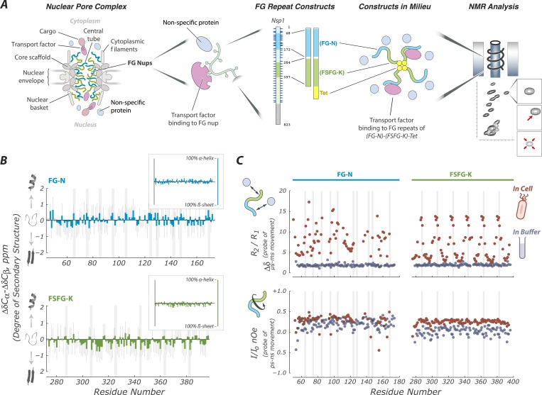
Figure 1.
Full legend
FG Nups are normally in a fully disordered and highly dynamic fluid state. (A) Our experimental approach includes key features of the NPC; a mixture of FG flavors, attachment at one end, and both specific (TF) and non-specific interactions with the cellular milieu. For example, our largest construct (FG-N-FSFG-K-tet) contains two fragments from Nsp1 (FG-N, turquoise; FSFG-K, green; full-length Nsp1 also shown with residue numbering), a separator (white) and the tetramerization domain of p53 (yellow). NMR analysis is performed on this construct and its variants, in milieu of various types; changes in position or intensity of peaks (bottom right) indicate changes in structure or interactions of the FG motifs. (B) Deviations of chemical shift values in cell (Escherichia coli) from predicted (colored bars) showing that FG-N and FSFG-K fragments are fully disordered, with no propensity for secondary structure. Also shown are standard errors of the mean (gray bars) and positions of FG motifs in the sequence (gray columns). Chemical shift values expected for an α-helix or β-sheet are approximately −4 and +4 ppm, respectively, as shown in the small insets. (C) The FG-N and FSFG-K constructs show significant interactions with the cellular milieu and exhibit very rapid motions. The upper panels show the ratio of R2/R1 indicative of overall motion and effects of multiple environments (chemical exchange) in cell (E. coli), compared to buffer A. The heteronuclear nuclear overhauser effect (nOe) is shown in lower panels, indicative of backbone motions. Gray columns indicate the locations of the FG motifs. Full experimental details and interpretations are available in ‘Materials and methods’ and Figure 1—figure supplements 1–4. DOI: http://dx.doi.org/10.7554/eLife.10027.003
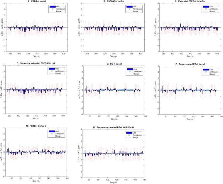
Figure 1—figure supplement 1.
Full legend
Lack of indicated secondary structure in FG Nup constructs (panels A–H, text). The most generally accepted indicator of propensity for secondary structure is the differences in deviations of 13C shifts of Cα and Cβ's (e.g., Spera and Bax, 1991; Wishart and Sykes, 1994; Wishart and Case, 2001, illustrated in e.g., Fushman et al., 1998; McDonnell et al., 1999; Mittag and Forman-Kay, 2007), with α-helical segments showing a value of ∼ +4, and β sheets a value of ∼ −4. However, the standard deviations of the underlying reference values have not been widely available until recently (Tamiola et al., 2010). In the above panels, the observed values of the difference of deviations for each residues is shown in blue (-●) while the range of the calculated standard errors is shown in red ( -- ). Values which are ambiguous or missing because of overlaps are indicated by an open circle ( O ). Panels are (A) FSFG-K in cell; (B) FSFG-K in buffer A; (C) same as B but those ‘degenerate’ sequences otherwise unassigned, are duplicated at the additional positions; for example, in this case the sequence surrounding P397 was most similar to that of P282 (ATSKPAFSF and ATSKPALEH) and the P282 values are used at position P397 for the 13Cα and 13Cβ assignments; (D) assignments of B extended; (E) FG-N in cell; (F) sequence duplicated values of E; (G) FG-N in buffer D; (H) Sequence extended values of set G. The data presented indicate no significant runs of deviation of the Δδ shift value, and none exceeding the standard deviations of the measurements. All conclusions in the paper are based on specific assignment, or grouped assignments in the case of overlaps and extended assignment, and do not rely on specific assignment in ambiguous/overlapped cases. When in cell assignments were ambiguous, and in buffer assignments were consistent with in cell observation, the in buffer assignments for specific residues were used. DOI: http://dx.doi.org/10.7554/eLife.10027.004
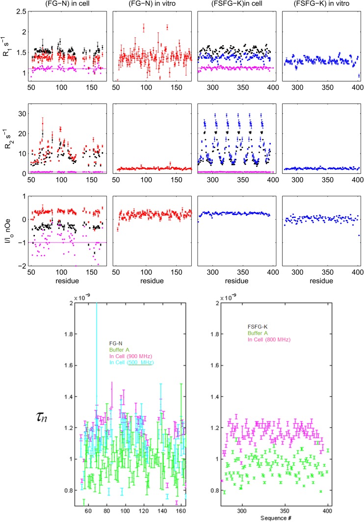
Figure 1—figure supplement 2.
Full legend
Details of NMR relaxation data and derived correlation times. Upper: 15N[1H] relaxation data for FG-N and FSFG-K in respectively E. coli cell, and in buffer A (‘in vitro’). Measurements were at 900 MHz (red), 800 MHz (blue), and 500 MHz (black). For those with dual field measurements, the ratio at the two fields is shown in purple. The nOe is used to analyze rapid backbone motion in the panel below, and data will be further examined in future work. Lower: derived rotational correlation times, τn, for the N-H bond vector from the heteronuclear nOe. Data indicate that the field dependence of the observed nOe for FG-N in cell (red and black in low left above) is almost solely from the expected field dependence of the expected correlation, since the derived τn's are very similar (purple and cyan in left below). The in buffer average values, ∼0.9 ns, are similar for the two constructs, consistent with their similar chemical shift and dynamic light scattering properties (Figure 1, Figure 4G). The measured errors for the FG-N are greater than those of the FSFG-K set, predominantly because accurate estimates of peak heights are more difficult in the presence of overlaps in the spectra. Although there appears to be some significant sequence dependence in the data for FSFG-K in-buffer (green, right), the S/N in the FG-N case obscures whether this is present there also. Sequence dependence of the τn may arise from local restriction by immediately adjacent side chains. Values of τn in cell are increased modestly (to ∼1.17 ns) for both constructs and are assumed to reflect partial restriction of fast internal motions (Neuhaus and Williamson, 2000). DOI: http://dx.doi.org/10.7554/eLife.10027.005
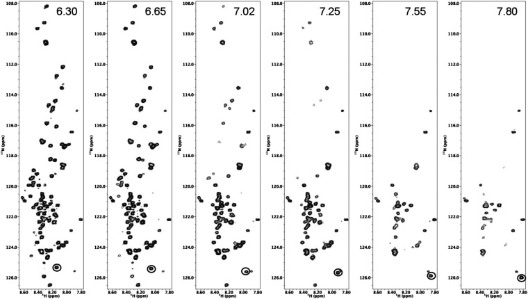
Figure 1—figure supplement 3.
Full legend
HSQC spectra of FSFG-K construct in buffer A vary with pH. HSQC spectra of FSFG-K in buffer A adjusted to the indicated pH's. In buffer, fast solvent exchange of significant number of peaks is apparent as expected (Croke et al., 2008, 2011; Burz et al., 2012). The His6 tag signal titrates with the pH change and is circled at the lower right of each panel. DOI: http://dx.doi.org/10.7554/eLife.10027.006
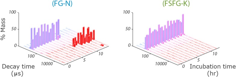
Figure 1—figure supplement 4.
Full legend
Stability of FG Nup constructs by DLS. Stability of size for FG-N (left) and FSFG-K (right) in buffer A by dynamic light scattering. The raw decay times are used so no interpretive models are applied. FG-N shows a sharp transition to a gel-like state after about 7 hr. Protein concentrations 8 mg/ml. For detailed methods see ‘Dynamic Light Scattering’. DOI: http://dx.doi.org/10.7554/eLife.10027.007
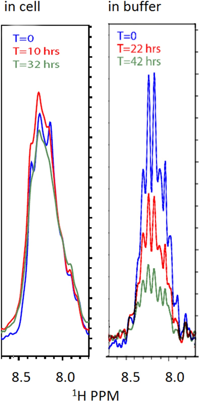
Figure 1—figure supplement 5.
Full legend
Stability of the FG-N construct in cell (left) and in buffer (right) by NMR. Left panel: freshly made cells (grown as ‘Materials and methods’); induced at OD 0.8 for 200 m were spun and resuspended in buffer E. Right panel: purified and concentrated FG-N was buffer exchanged directly from 8 M urea to buffer A. All spectrometer conditions remained constant between scans. Samples were contained in 5 mm Shigemi NMR tubes. The spectra are 1-D traces from the first block of HSQC spectra initiated at the listed times. DOI: http://dx.doi.org/10.7554/eLife.10027.008
Show all 13 figures
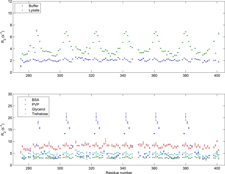
Figure 1—figure supplement 6.
Full legend
Transverse relaxation of FSFG-K in multiple environments. All R2 data were collected at 25°C with 140 μM 15N-labeled FSFG-K in buffer A with the following delays: 0, 32.64, 65.28, 97.92, 130.56, 163.20, 195.84, 228.48, 261.12, and 326.40 ms. The concentrations of the different additives are as follows: E. coli lysate (Wang et al., 2011; Latham and Kay, 2014) 230 mg/ml; BSA 205 mg/ml Glycerol (Li et al., 2009; Wang et al., 2011) 60% wt/wt; PVP-10 (Li et al., 2008, 2009) 111 mg/ml; Trehalose (Lins et al., 2004; Chakrabortee et al., 2010) 154 mg/ml. Sequence-dependent fluctuation is seen only with lysate or with BSA, indicating non-specific rapid interactions, in contrast to changes of viscosity (Serber et al., 2005; Li et al., 2009; Wang et al., 2011), solvation (Lim and Deng, 2009; Xue and Skrynnikov, 2011; Kim et al., 2013), or packing (Li et al., 2009; Uversky, 2009; Milles et al., 2013). The E. coli lysate concentration is comparable to the estimated in vivo total concentration (Cayley et al., 1991). Interaction with BSA is consistent with previous observations (Parks et al., 1983; Higuchi et al., 1994; Clarkson et al., 1996; Fielding et al., 2005; Simard et al., 2006). DOI: http://dx.doi.org/10.7554/eLife.10027.009
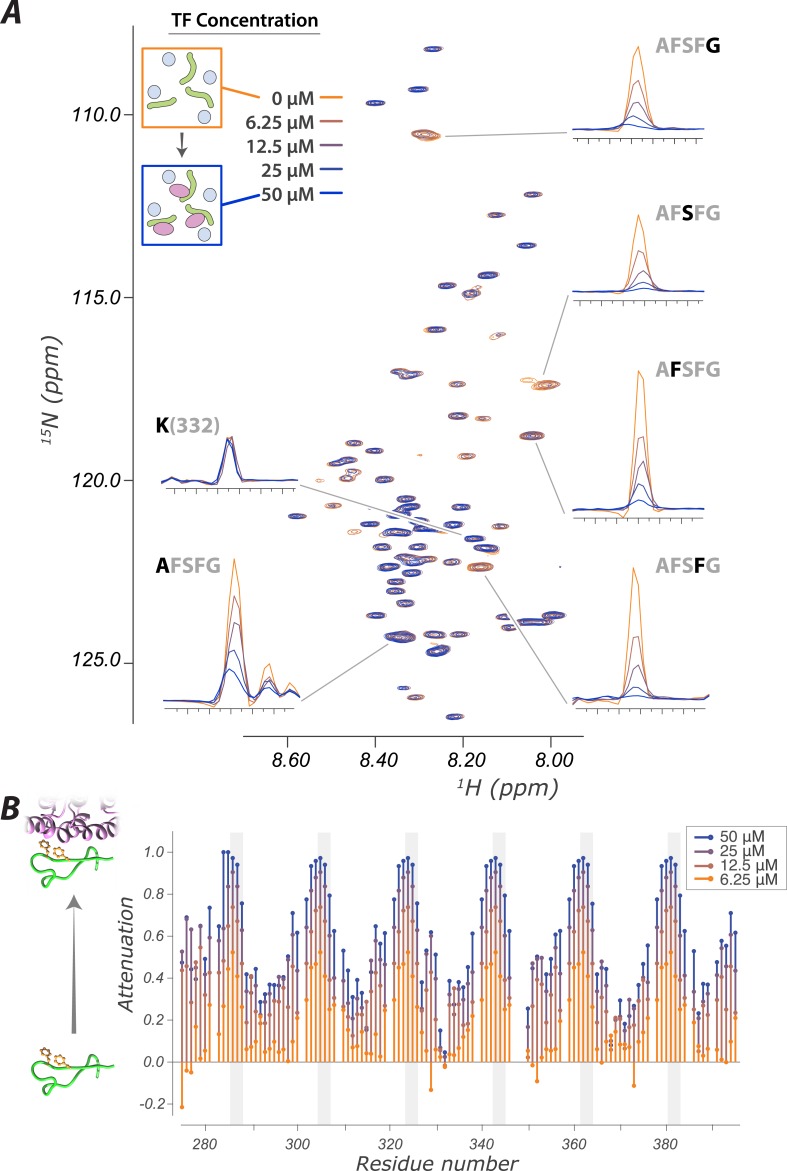
Figure 2.
Full legend
The interaction of FG Nups with the transport factor Kap95 is specific to the FG sites and leaves the spacers highly mobile (A) Overlay of spectra with varying concentrations of Kap95 (0, 6.125, 12.5, 25, 50 μM) in the presence of [U-15N] FSFG-K (25 μM) in Xenopus egg extract, showing only the FG motifs strongly interact with the TF. Several peaks represent overlapping similar sequences with indistinguishable attenuation. (B) Superimposed values of attenuation ((1 − I)/Io) on addition of Kap95 across the sequence; increased attenuation indicates a stronger interaction propensity for that residue (positions of FG motifs in the sequence are indicated by gray columns). DOI: http://dx.doi.org/10.7554/eLife.10027.010
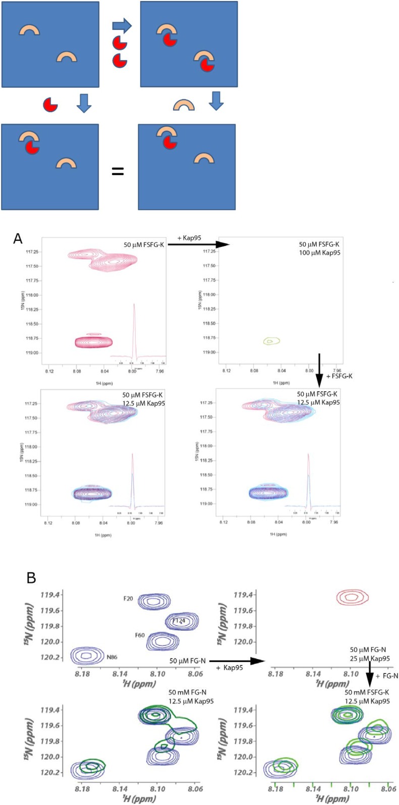
Figure 2—figure supplement 1.
Full legend
Reversibility of TF/FG Nup interactions. For each of FSFG-GK and FG-N's interactions with Kap95, we demonstrated reversibility as follows. The spectra below can be cartooned as arising from the following, where the upper left panel represents the solution of an FG Nup. Addition of TFs (red) at high concentration forms a substantial amount of complex (upper right). Addition of more FG Nup at the same concentration as upper left results in reversibility of a portion of the complex formed, and spectra identical to those formed by direct addition of TF in smaller amounts (lower panels). If the complex was irreversible, then the dilution from the right upper complex, would lead to a summation of the upper two figures for the right lower, and would not show the intermediate chemical shifts seen. (A) HSQC spectra of complexes of FSFG-K and Kap95, illustrating the formation of the same complex (lower spectra) by either admixture of the components (lower left) or dilution of the TF concentration from a higher value to the final by addition of FSFG-K (lower right). At upper left, the region of the S, F residues of FSFG-K 50 μM is shown in the absence of Kap95 in buffer C. On makeup to 100 μM Kap95, the resulting spectrum is at the upper right. Dilution with additional FSFG-K produces the spectra at the lower right. The lower spectra superimpose the starting spectrum (upper left, red) and the complex spectrum (azure). (B) HSQC spectra of complexes of FG-N and Kap95 components (lower left) or dilution of the TF concentration from a higher value to the final by addition of FG-N (lower right) in buffer A. At upper left, the region of the N and F residues of FG-N 50 μM is shown in the absence of Kap95. On addition of Kap95 to 0.5 molar equivalence (25 μM), the resulting spectrum is at the upper right. Dilution with additional FG-N produces the spectra at the lower right. The lower right spectra superimpose the starting spectrum (upper left, blue) and the complex spectrum (green). DOI: http://dx.doi.org/10.7554/eLife.10027.011
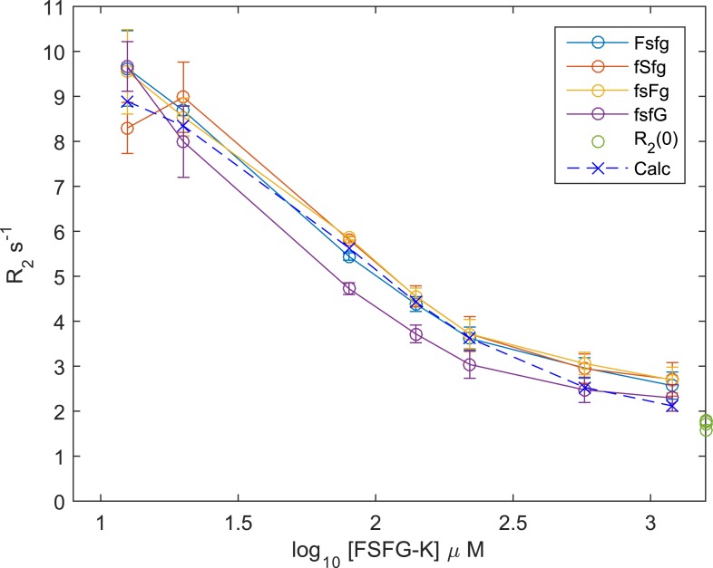
Figure 2—figure supplement 2.
Full legend
15N R2 titration of Kap95 and [15N]FSFG-K. Observed values of 15N R2 for FSFG-K at various concentrations, in the presence of 20 μM Kap95. Experimental details in ‘NMR analysis’ in ‘Materials and methods’. The 15N R2 values and their standard errors of the mean are plotted vs the log10 of the 15N FGFG-K concentration all in the presence of 20 μM Kap95 for the residue types of the FSFG motif. The values of 15N R2 are shown in green at the extreme right axis for free FSFG-K. The derived value fitted for the equation of Section S2.3 is shown as the dotted line with ‘X’ marks. The fitted values of Kd and Rb for the derived line are 36.1 μM and 25.5 s−1. The apparent Kd is then equivalent to 216 μM for each FSFG site in FSFG-K. Lines connect the observed or calculated values. DOI: http://dx.doi.org/10.7554/eLife.10027.012
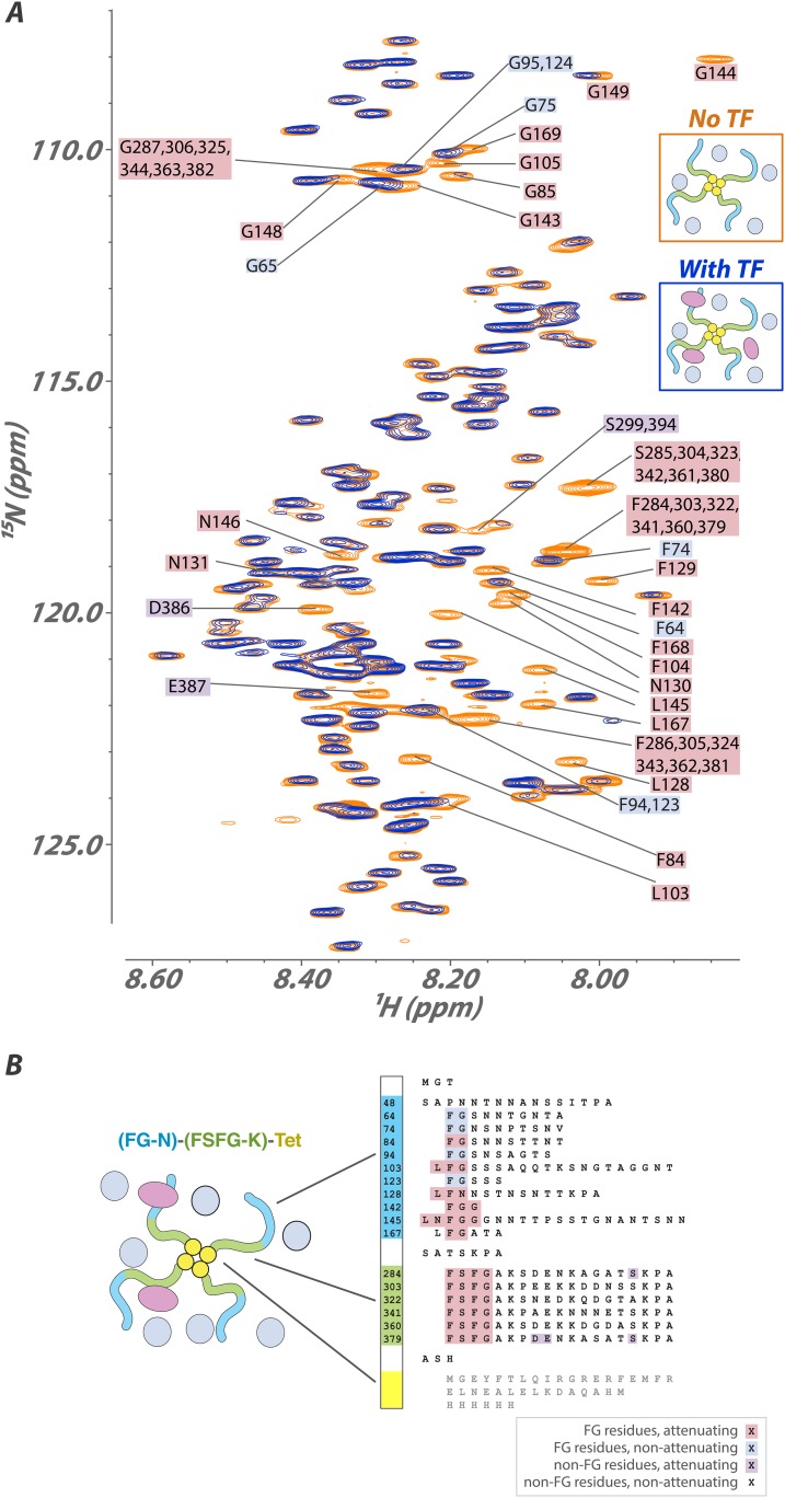
Figure 3.
Full legend
Interaction of a high molecular-weight tetramerized hybrid construct indicates that the binding mechanism is robust to changes in FG type, packing, and environment. (A) 1H[15N] Heteronuclear Single Quantum Coherence (HSQC) spectra of the FG-N-FSFG-K-tet sequence (Tables 1 and 2) (MW 120 kDa) in the unbound (orange) and Kap95-bound (blue) states with Xenopus egg extract as the milieu. Peaks that undergo signal attenuation in the Kap95-bound spectrum indicate binding of those residues to Kap95. Typically, we observe that it is the FG residues (pink boxes), but not intervening spacer residues (no boxes), that attenuate upon binding to the transport factor Kap95; however, there are four FG repeats that are minimally attenuated (blue boxes), and a small number of spacer residues that are attenuated (purple boxes). (B) Schematic of the construct used, labeled as in (A). Very small chemical shift changes are observable for a few sites, consistent with rapid exchange into multiple inhomogeneous sites. DOI: http://dx.doi.org/10.7554/eLife.10027.013
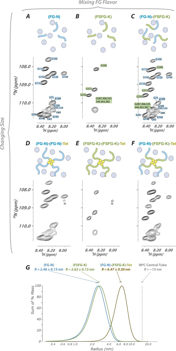
Figure 4.
Full legend
FG constructs show no significant FG–FG interactions, and remain fully disordered even when tethered at one end and packed at high density. (A–F) We developed a wide range of constructs containing fragments from Nsp1 (turquoise and green, numbering in Figure 1) and the small tetramerization domain from p53 (yellow). Schematics illustrate the different constructs as studied within the cellular milieu (blue circles) which vary both in total molecular weight, crowding of the FG repeats via tetramerization, and composition of FG fragments (homotypic or heterotypic). Below each schematic is the HSQC spectrum of the glycine region of the represented construct observed in cell (E. coli). All constructs are tabulated in Table 1. ‘M’ identifies apparent 15N-labeled metabolites which are variable in intensity from preparation to preparation. (G) Dynamic light scattering of three representative constructs in buffer, showing homogeneity and consistency with expected size of constructs. DOI: http://dx.doi.org/10.7554/eLife.10027.014
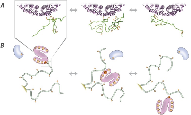
Figure 5.
Full legend
Molecular description of the nuclear transport mechanism. Specific conclusions from our results, as discussed in the main text, are illustrated with (A) details from a docked molecular simulation of the TF Kap95 (purple) and the FG repeat region FSFG-K, rendered in Chimera (see ‘Materials and methods’), and (B) a diagrammatic view of the molecules involved. FG repeat (green; an ensemble of disordered conformers is illustrated in A). Phe (orange), Phe-binding pockets (B, orange circles), TF (pink), non-specific macromolecule (B, blue). DOI: http://dx.doi.org/10.7554/eLife.10027.015
References139
- 1.
Acuner Ozbabacan SE, Engin HB, Gursoy A, Keskin O. Transient protein-protein interactions. Protein Engineering, Design & Selection. 2011;24:635-648.
Not in our library10.1093/protein/gzr025 (opens in a new tab)
- 2.
Adams RL, Wente SR. Uncovering nuclear pore complexity with innovation. Cell. 2013;152:1218-1221.
Not in our library10.1016/j.cell.2013.02.042 (opens in a new tab)
- 3.
Ader C, Frey S, Maas W, Schmidt HB, Görlich D, Baldus M. Amyloid-like interactions within nucleoporin FG hydrogels. Proceedings of the National Academy of Sciences of USA. 2010;107:6281-6285.
Not in our library10.1073/pnas.0910163107 (opens in a new tab)
- 4.
Akey CW, Radermacher M. Architecture of the Xenopus nuclear pore complex revealed by three-dimensional cryo-electron microscopy. Journal of Cell Biology. 1993;122:1-19.
Not in our library10.1083/jcb.122.1.1 (opens in a new tab)
- 5.
Alber F, Dokudovskaya S, Veenhoff LM, Zhang W, Kipper J, Devos D, Suprapto A, Karni-Schmidt O, Williams R, Chait BT, Sali A, Rout MP. The molecular architecture of the nuclear pore complex. Nature. 2007;450:695-701.
Not in our library10.1038/nature06405 (opens in a new tab)
- 6.
Atkinson CE, Mattheyses AL, Kampmann M, Simon SM. Conserved spatial organization of FG domains in the nuclear pore complex. Biophysical Journal. 2013;104:37-50.
Not in our library10.1016/j.bpj.2012.11.3823 (opens in a new tab)
- 7.
Barbato G, Ikura M, Kay LE, Pastor RW, Bax A. Backbone dynamics of calmodulin studied by 15N relaxation using inverse detected two-dimensional NMR spectroscopy: the central helix is flexible. Biochemistry. 1992;31:5269-5278.
Not in our library10.1021/bi00138a005 (opens in a new tab)
- 8.
Bayliss R, Corbett AH, Stewart M. The molecular mechanism of transport of macromolecules through nuclear pore complexes. Traffic. 2000;1:448-456.
Not in our library10.1034/j.1600-0854.2000.010602.x (opens in a new tab)
- 9.
Bayliss R, Littlewood T, Strawn LA, Wente SR, Stewart M. GLFG and FxFG nucleoporins bind to overlapping sites on importin-beta. The Journal of Biological Chemistry. 2002;277:50597-50606.
Not in our library10.1074/jbc.m209037200 (opens in a new tab)
- 10.
Bednenko J, Cingolani G, Gerace L. Importin beta contains a COOH-terminal nucleoporin binding region important for nuclear transport. Journal of Cell Biology. 2003;162:391-401.
Not in our library10.1083/jcb.200303085 (opens in a new tab)
- 11.
Berne BJ, Pecora F. Dynamic light scattering with applications to chemistry, biology, and physics. 2000.
Not in our library
- 12.
Buevich AV, Shinde UP, Inouye M, Baum J. Backbone dynamics of the natively unfolded pro-peptide of subtilisin by heteronuclear NMR relaxation studies. Journal of Biomolecular NMR. 2001;20:233-249.
Not in our library10.1023/a:1011243116136 (opens in a new tab)
- 13.
Burns LT, Wente SR. Trafficking to uncharted territory of the nuclear envelope. Current Opinion in Cell Biology. 2012;24:341-349.
Not in our library10.1016/j.ceb.2012.01.009 (opens in a new tab)
- 14.
Burz DS, Cowburn D, Dutta K, Shekhtman A, Bertini I. NMR of biomolecules. 2012.
Not in our library
- 15.
Burz DS, Dutta K, Cowburn D, Shekhtman A. In-cell NMR for protein-protein interactions (STINT-NMR). Nature Protocols. 2006a;1:146-152.
Not in our library10.1038/nprot.2006.23 (opens in a new tab)
- 16.
Burz DS, Dutta K, Cowburn D, Shekhtman A. Mapping structural interactions using in-cell NMR spectroscopy (STINT-NMR). Nature Methods. 2006b;3:91-93.
Not in our library10.1038/nmeth851 (opens in a new tab)
- 17.
Cantor C, Schimmel P. Biophysical chemistry, part III: the behavior of biological macromolecules. 1980;1133.
Not in our library
- 18.
Cavanagh J, Fairbrother WJ, Palmer AGI, Rance M, Skelton NJ. Protein NMR spectroscopy: principles and practice. 2007.
Not in our library
- 19.
Cayley S, Lewis BA, Guttman HJ, Record MT. Characterization of the cytoplasm of Escherichia coli K-12 as a function of external osmolarity. Implications for protein-DNA interactions in vivo. Journal of Molecular Biology. 1991;222:281-300.
Not in our library10.1016/0022-2836(91)90212-o (opens in a new tab)
- 20.
Chakrabortee S, Meersman F, Kaminski Schierle GS, Bertoncini CW, McGee B, Kaminski CF, Tunnacliffe A. Catalytic and chaperone-like functions in an intrinsically disordered protein associated with desiccation tolerance. Proceedings of the National Academy of Sciences of USA. 2010;107:16084-16089.
Not in our library10.1073/pnas.1006276107 (opens in a new tab)
- 21.
Clackson T, Wells JA. A hot spot of binding energy in a hormone-receptor interface. Science. 1995;267:383-386.
Not in our library10.1126/science.7529940 (opens in a new tab)
- 22.
Clarkson WD, Kent HM, Stewart M. Separate binding sites on nuclear transport factor 2 (NTF2) for GDP-Ran and the phenylalanine-rich repeat regions of nucleoporins p62 and Nsp1p. Journal of Molecular Biology. 1996;263:517-524.
Not in our library10.1006/jmbi.1996.0594 (opens in a new tab)
- 23.
Croke RL, Patil SM, Quevreaux J, Kendall DA, Alexandrescu AT. NMR determination of pKa values in alpha-synuclein. Protein Science. 2011;20:256-269.
Not in our library10.1002/pro.556 (opens in a new tab)
- 24.
Croke RL, Sallum CO, Watson E, Watt ED, Alexandrescu AT. Hydrogen exchange of monomeric alpha-synuclein shows unfolded structure persists at physiological temperature and is independent of molecular crowding in Escherichia coli. Protein Science. 2008;17:1434-1445.
Not in our library10.1110/ps.033803.107 (opens in a new tab)
- 25.
Dabauvalle MC, Loos K, Merkert H, Scheer U. Spontaneous assembly of pore complex-containing membranes (‘annulate lamellae’) in Xenopus egg extract in the absence of chromatin. Journal of Cell Biology. 1991;112:1073-1082.
Not in our library10.1083/jcb.112.6.1073 (opens in a new tab)
- 26.
- 27.
Dixon SE, Bhatti MM, Uversky VN, Dunker AK, Sullivan WJ. Regions of intrinsic disorder help identify a novel nuclear localization signal in Toxoplasma gondii histone acetyltransferase TgGCN5-B. Molecular and Biochemical Parasitology. 2011;175:192-195.
Not in our library10.1016/j.molbiopara.2010.10.009 (opens in a new tab)
- 28.
Dominguez C, Boelens R, Bonvin AM. HADDOCK: a protein-protein docking approach based on biochemical or biophysical information. Journal of the American Chemical Society. 2003;125:1731-1737.
Not in our library10.1021/ja026939x (opens in a new tab)
- 29.
Eisele NB, Frey S, Piehler J, Görlich D, Richter RP. Ultrathin nucleoporin phenylalanine-glycine repeat films and their interaction with nuclear transport receptors. EMBO Reports. 2010;11:366-372.
Not in our library10.1038/embor.2010.34 (opens in a new tab)
- 30.
Eliezer D. Biophysical characterization of intrinsically disordered proteins. Current Opinion in Structural Biology. 2009;19:23-30.
Not in our library10.1016/j.sbi.2008.12.004 (opens in a new tab)
- 31.
Feeney J, Batchelor J, Albrand J, Roberts G. The effects of intermediate exchange processes on the estimation of equilibrium constants by NMR. Journal of Magnetic Resonance. 1979;33:519-529.
Not in our library
- 32.
Ferrage F, Cowburn D, Ghose R. Accurate sampling of high-frequency motions in proteins by steady-state (15)N-{(1)H} nuclear overhauser effect measurements in the presence of cross-correlated relaxation. Journal of the American Chemical Society. 2009;131:6048-6049.
Not in our library10.1021/ja809526q (opens in a new tab)
- 33.
Ferrage F, Pelupessy P, Cowburn D, Bodenhausen G. Protein backbone dynamics through 13C′-13Calpha cross-relaxation in NMR spectroscopy. Journal of the American Chemical Society. 2006;128:11072-11078.
Not in our library10.1021/ja0600577 (opens in a new tab)
- 34.
Ferrage F, Reichel A, Battacharya S, Cowburn D, Ghose R. On the measurement of (1)(5)N-{(1)H} nuclear Overhauser effects. 2. Effects of the saturation scheme and water signal suppression. Journal of Magnetic Resonance. 2010;207:294-303.
Not in our library10.1016/j.jmr.2010.09.014 (opens in a new tab)
- 35.
Fielding L, Rutherford S, Fletcher D. Determination of protein-ligand binding affinity by NMR: observations from serum albumin model systems. Magnetic Resonance in Chemistry. 2005;43:463-470.
Not in our library10.1002/mrc.1574 (opens in a new tab)
- 36.
Frey S, Görlich D. A saturated FG-repeat hydrogel can reproduce the permeability properties of nuclear pore complexes. Cell. 2007;130:512-523.
Not in our library10.1016/j.cell.2007.06.024 (opens in a new tab)
- 37.
Frey S, Görlich D. FG/FxFG as well as GLFG repeats form a selective permeability barrier with self-healing properties. The EMBO Journal. 2009;28:2554-2567.
Not in our library10.1038/emboj.2009.199 (opens in a new tab)
- 38.
Frey S, Richter RP, Görlich D. FG-rich repeats of nuclear pore proteins form a three-dimensional meshwork with hydrogel-like properties. Science. 2006;314:815-817.
Not in our library10.1126/science.1132516 (opens in a new tab)
- 39.
Funasaka T, Balan V, Raz A, Wong RW. Nucleoporin Nup98 mediates galectin-3 nuclear-cytoplasmic trafficking. Biochemical and Biophysical Research Communications. 2013;434:155-161.
Not in our library10.1016/j.bbrc.2013.03.052 (opens in a new tab)
- 40.
Fushman D, Cahill S, Cowburn D. The main-chain dynamics of the dynamin pleckstrin homology (PH) domain in solution: analysis of 15N relaxation with monomer/dimer equilibration. Journal of Molecular Biology. 1997;266:173-194.
Not in our library10.1006/jmbi.1996.0771 (opens in a new tab)
- 41.
Fushman D, Najmabadi-Haske T, Cahill S, Zheng J, LeVine H, Cowburn D. The solution structure and dynamics of the pleckstrin homology domain of G protein-coupled receptor kinase 2 (beta-adrenergic receptor kinase 1). A binding partner of Gbetagamma subunits. The Journal of Biological Chemistry. 1998;273:2835-2843.
Not in our library10.1074/jbc.273.5.2835 (opens in a new tab)
- 42.
Fuxreiter M, Tompa P. Fuzzy complexes: a more stochastic view of protein function. Advances in Experimental Medicine and Biology. 2012;725:1-14.
Not in our library10.1007/978-1-4614-0659-4_1 (opens in a new tab)
- 43.
Gamini R, Han W, Stone JE, Schulten K. Assembly of Nsp1 nucleoporins provides insight into nuclear pore complex gating. PLOS Computational Biology. 2014;10:e1003488.
Not in our library10.1371/journal.pcbi.1003488 (opens in a new tab)
- 44.
Gant TM, Goldberg MW, Allen TD. Nuclear envelope and nuclear pore assembly: analysis of assembly intermediates by electron microscopy. Current Opinion in Cell Biology. 1998;10:409-415.
Not in our library10.1016/s0955-0674(98)80018-5 (opens in a new tab)
- 45.
Goldberg MW, Allen TD. The nuclear pore complex and lamina: three-dimensional structures and interactions determined by field emission in-lens scanning electron microscopy. Journal of Molecular Biology. 1996;257:848-865.
Not in our library10.1006/jmbi.1996.0206 (opens in a new tab)
- 46.
Goldflam M, Tarragó T, Gairí M, Giralt E, Shekhtman A, Burz DS. Protein NMR techniques. Methods in molecular biology. 2012.
Not in our library
- 47.
Goldschmidt L, Teng PK, Riek R, Eisenberg D. Identifying the amylome, proteins capable of forming amyloid-like fibrils. Proceedings of the National Academy of Sciences of USA. 2010;107:3487-3492.
Not in our library10.1073/pnas.0915166107 (opens in a new tab)
- 48.
Han TW, Kato M, Xie S, Wu LC, Mirzaei H, Pei J, Chen M, Xie Y, Allen J, Xiao G, McKnight SL. Cell-free formation of RNA granules: bound RNAs identify features and components of cellular assemblies. Cell. 2012;149:768-779.
Not in our library10.1016/j.cell.2012.04.016 (opens in a new tab)
- 49.
Hanlon AD, Larkin MI, Reddick RM. Free-solution, label-free protein-protein interactions characterized by dynamic light scattering. Biophysical Journal. 2010;98:297-304.
Not in our library10.1016/j.bpj.2009.09.061 (opens in a new tab)
- 50.
Higuchi A, Hara M, Horiuchi T, Nakagawa T. Optical resolution of amino-acids by ultrafiltration membranes containing serum-albumin. Journal of Membrane Science. 1994;93:157-164.
Not in our library10.1016/0376-7388(94)80004-9 (opens in a new tab)
- 51.
Hulsmann BB, Labokha AA, Görlich D. The permeability of reconstituted nuclear pores provides direct evidence for the selective phase model. Cell. 2012;150:738-751.
Not in our library10.1016/j.cell.2012.07.019 (opens in a new tab)
- 52.
Isgro TA, Schulten K. Binding dynamics of isolated nucleoporin repeat regions to importin-beta. Structure. 2005;13:1869-1879.
Not in our library10.1016/j.str.2005.09.007 (opens in a new tab)
- 53.
Isgro TA, Schulten K. Cse1p-binding dynamics reveal a binding pattern for FG-repeat nucleoporins on transport receptors. Structure. 2007;15:977-991.
Not in our library10.1016/j.str.2007.06.011 (opens in a new tab)
- 54.
Jovanovic-Talisman T, Tetenbaum-Novatt J, McKenney AS, Zilman A, Peters R, Rout MP, Chait BT. Artificial nanopores that mimic the transport selectivity of the nuclear pore complex. Nature. 2009;457:1023-1027.
Not in our library10.1038/nature07600 (opens in a new tab)
- 55.
Kalinina J, Dutta K, Ilghari D, Beenken A, Goetz R, Eliseenkova AV, Cowburn D, Mohammadi M. The alternatively spliced acid box region plays a key role in FGF receptor autoinhibition. Structure. 2012;20:77-88.
Not in our library10.1016/j.str.2011.10.022 (opens in a new tab)
- 56.
Kapinos LE, Schoch RL, Wagner RS, Schleicher KD, Lim RY. Karyopherin-centric control of nuclear pores based on molecular occupancy and kinetic analysis of multivalent binding with FG nucleoporins. Biophysical Journal. 2014;106:1751-1762.
Not in our library10.1016/j.bpj.2014.02.021 (opens in a new tab)
- 57.
Kato M, Han TW, Xie S, Shi K, Du X, Wu LC, Mirzaei H, Goldsmith EJ, Longgood J, Pei J, Grishin NV, Frantz DE, Schneider JW, Chen S, Li L, Sawaya MR, Eisenberg D, Tycko R, McKnight SL. Cell-free formation of RNA granules: low complexity sequence domains form dynamic fibers within hydrogels. Cell. 2012;149:753-767.
Not in our library10.1016/j.cell.2012.04.017 (opens in a new tab)
- 58.
Kim S, Wu KP, Baum J. Fast hydrogen exchange affects (1)(5)N relaxation measurements in intrinsically disordered proteins. Journal of Biomolecular NMR. 2013;55:249-256.
Not in our library10.1007/s10858-013-9706-1 (opens in a new tab)
- 59.
Kiseleva E, Allen TD, Rutherford S, Bucci M, Wente SR, Goldberg MW. Yeast nuclear pore complexes have a cytoplasmic ring and internal filaments. Journal of Structural Biology. 2004;145:272-288.
Not in our library10.1016/j.jsb.2003.11.010 (opens in a new tab)
- 60.
Kleckner IR, Foster MP. An introduction to NMR-based approaches for measuring protein dynamics. Biochimica et Biophysica Acta. 2011;1814:942-968.
Not in our library10.1016/j.bbapap.2010.10.012 (opens in a new tab)
- 61.
Koppel DE. Analysis of macromolecular polydispersity in intensity correlation spectroscopy: the method of cumulants. Journal of Chemical Physics. 1972;57:4814.
Not in our library10.1063/1.1678153 (opens in a new tab)
- 62.
- 63.
Kowalczyk SW, Kapinos L, Blosser TR, Magalhães T, van Nies P, Lim RY, Dekker C. Single-molecule transport across an individual biomimetic nuclear pore complex. Nature Nanotechnology. 2011;6:433-438.
Not in our library10.1038/nnano.2011.88 (opens in a new tab)
- 64.
Laba JK, Steen A, Veenhoff LM. Traffic to the inner membrane of the nuclear envelope. Current Opinion in Cell Biology. 2014;28:36-45.
Not in our library10.1016/j.ceb.2014.01.006 (opens in a new tab)
- 65.
Labokha AA, Gradmann S, Frey S, Hülsmann BB, Urlaub H, Baldus M, Görlich D. Systematic analysis of barrier-forming FG hydrogels from Xenopus nuclear pore complexes. The EMBO Journal. 2013;32:204-218.
Not in our library10.1038/emboj.2012.302 (opens in a new tab)
- 66.
Latham MP, Kay LE. A similar in vitro and in cell lysate folding intermediate for the FF domain. Journal of Molecular Biology. 2014;426:3214-3220.
Not in our library10.1016/j.jmb.2014.07.019 (opens in a new tab)
- 67.
Lemak A, Gutmanas A, Chitayat S, Karra M, Farès C, Sunnerhagen M, Arrowsmith CH. A novel strategy for NMR resonance assignment and protein structure determination. Journal of Biomolecular NMR. 2011;49:27-38.
Not in our library10.1007/s10858-010-9458-0 (opens in a new tab)
- 68.
Lepre CA, Moore JM, Peng JW. Theory and applications of NMR-based screening in pharmaceutical research. Chemical Reviews. 2004;104:3641-3676.
Not in our library10.1021/cr030409h (opens in a new tab)
- 69.
Li C, Charlton LM, Lakkavaram A, Seagle C, Wang G, Young GB, Macdonald JM, Pielak GJ. Differential dynamical effects of macromolecular crowding on an intrinsically disordered protein and a globular protein: implications for in-cell NMR spectroscopy. Journal of the American Chemical Society. 2008;130:6310-6311.
Not in our library10.1021/ja801020z (opens in a new tab)
- 70.
Li C, Wang Y, Pielak GJ. Translational and rotational diffusion of a small globular protein under crowded conditions. The Journal of Physical Chemistry. B. 2009;113:13390-13392.
Not in our library10.1021/jp907744m (opens in a new tab)
- 71.
Libich DS, Fawzi NL, Ying J, Clore GM. Probing the transient dark state of substrate binding to GroEL by relaxation-based solution NMR. Proceedings of the National Academy of Sciences of USA. 2013;110:11361-11366.
Not in our library10.1073/pnas.1305715110 (opens in a new tab)
- 72.
Lim RY, Deng J. Interaction forces and reversible collapse of a polymer brush-gated nanopore. ACS Nano. 2009;3:2911-2918.
Not in our library10.1021/nn900152m (opens in a new tab)
- 73.
Lim RY, Fahrenkrog B, Köser J, Schwarz-Herion K, Deng J, Aebi U. Nanomechanical basis of selective gating by the nuclear pore complex. Science. 2007;318:640-643.
Not in our library10.1126/science.1145980 (opens in a new tab)
- 74.
Lim RY, Huang NP, Köser J, Deng J, Lau KH, Schwarz-Herion K, Fahrenkrog B, Aebi U. Flexible phenylalanine-glycine nucleoporins as entropic barriers to nucleocytoplasmic transport. Proceedings of the National Academy of Sciences of USA. 2006;103:9512-9517.
Not in our library10.1073/pnas.0603521103 (opens in a new tab)
- 75.
Lins RD, Pereira CS, Hünenberger PH. Trehalose-protein interaction in aqueous solution. Proteins. 2004;55:177-186.
Not in our library10.1002/prot.10632 (opens in a new tab)
- 76.
Liu SM, Stewart M. Structural basis for the high-affinity binding of nucleoporin Nup1p to the Saccharomyces cerevisiae importin-beta homologue, Kap95p. Journal of Molecular Biology. 2005;349:515-525.
Not in our library10.1016/j.jmb.2005.04.003 (opens in a new tab)
- 77.
- 78.
Luna RE, Akabayov SR, Ziarek JJ, Wagner G. Examining weak protein-protein interactions in start codon recognition via NMR spectroscopy. The FEBS Journal. 2014;281:1965-1973.
Not in our library10.1111/febs.12667 (opens in a new tab)
- 79.
Ma J, Goryaynov A, Sarma A, Yang W. Self-regulated viscous channel in the nuclear pore complex. Proceedings of the National Academy of Sciences of USA. 2012;109:7326-7331.
Not in our library10.1073/pnas.1201724109 (opens in a new tab)
- 80.
Magnusdottir A, Johansson I, Dahlgren LG, Nordlund P, Berglund H. Enabling IMAC purification of low abundance recombinant proteins from E. coli lysates. Nature Methods. 2009;6:477-478.
Not in our library10.1038/nmeth0709-477 (opens in a new tab)
- 81.
Mattheyses AL, Kampmann M, Atkinson CE, Simon SM. Fluorescence anisotropy reveals order and disorder of protein domains in the nuclear pore complex. Biophysical Journal. 2010;99:1706-1717.
Not in our library10.1016/j.bpj.2010.06.075 (opens in a new tab)
- 82.
McDonnell JM, Fushman D, Milliman CL, Korsmeyer SJ, Cowburn D. Solution structure of the proapoptotic molecule BID: a structural basis for apoptotic agonists and antagonists. Cell. 1999;96:625-634.
Not in our library10.1016/s0092-8674(00)80573-5 (opens in a new tab)
- 83.
Milles S, Huy Bui K, Koehler C, Eltsov M, Beck M, Lemke EA. Facilitated aggregation of FG nucleoporins under molecular crowding conditions. EMBO Reports. 2013;14:178-183.
Not in our library10.1038/embor.2012.204 (opens in a new tab)
- 84.
Mittag T, Forman-Kay JD. Atomic-level characterization of disordered protein ensembles. Current Opinion in Structural Biology. 2007;17:3-14.
Not in our library10.1016/j.sbi.2007.01.009 (opens in a new tab)
- 85.
Morrison J, Yang JC, Stewart M, Neuhaus D. Solution NMR study of the interaction between NTF2 and nucleoporin FxFG repeats. Journal of Molecular Biology. 2003;333:587-603.
Not in our library10.1016/j.jmb.2003.08.050 (opens in a new tab)
- 86.
- 87.
Muralidharan V, Dutta K, Cho J, Vila-Perello M, Raleigh DP, Cowburn D, Muir TW. Solution structure and folding characteristics of the C-terminal SH3 domain of c-Crk-II. Biochemistry. 2006;45:8874-8884.
Not in our library10.1021/bi060590z (opens in a new tab)
- 88.
Neuhaus D, Williamson MP. The nuclear overhauser effect in structural and conformational analysis. 2000.
Not in our library
- 89.
Nicholas MP, Eryilmaz E, Ferrage F, Cowburn D, Ghose R. Nuclear spin relaxation in isotropic and anisotropic media. Progress in nuclear magnetic resonance spectroscopy. 2010;57:111-158.
Not in our library10.1016/j.pnmrs.2010.04.003 (opens in a new tab)
- 90.
Parks JS, Cistola DP, Small DM, Hamilton JA. Interactions of the carboxyl group of oleic acid with bovine serum albumin: a 13C NMR study. The Journal of Biological Chemistry. 1983;258:9262-9269.
Not in our library
- 91.
Patel SS, Belmont BJ, Sante JM, Rexach MF. Natively unfolded nucleoporins gate protein diffusion across the nuclear pore complex. Cell. 2007;129:83-96.
Not in our library10.1016/j.cell.2007.01.044 (opens in a new tab)
- 92.
Peters R. Translocation through the nuclear pore complex: selectivity and speed by reduction-of-dimensionality. Traffic. 2005;6:421-427.
Not in our library10.1111/j.1600-0854.2005.00287.x (opens in a new tab)
- 93.
Phillip Y, Schreiber G. Formation of protein complexes in crowded environments–from in vitro to in vivo. FEBS Letters. 2013;587:1046-1052.
Not in our library10.1016/j.febslet.2013.01.007 (opens in a new tab)
- 94.
Poon GM, Brokx RD, Sung M, Gariépy J. Tandem dimerization of the human p53 tetramerization domain stabilizes a primary dimer intermediate and dramatically enhances its oligomeric stability. Journal of Molecular Biology. 2007;365:1217-1231.
Not in our library10.1016/j.jmb.2006.10.051 (opens in a new tab)
- 95.
Rout MP, Aitchison JD, Magnasco MO, Chait BT. Virtual gating and nuclear transport: the hole picture. Trends in Cell Biology. 2003;13:622-628.
Not in our library10.1016/j.tcb.2003.10.007 (opens in a new tab)
- 96.
Rout MP, Aitchison JD, Suprapto A, Hjertaas K, Zhao Y, Chait BT. The yeast nuclear pore complex: composition, architecture, and transport mechanism. Journal of Cell Biology. 2000;148:635-651.
Not in our library10.1083/jcb.148.4.635 (opens in a new tab)
- 97.
Sattler M, Schleucher J, Griesinger C. Heteronuclear multidimensional NMR experiments for the structure determination of proteins in solution employing pulsed field gradients. Progress in nuclear magnetic resonance spectroscopy. 1999;34:93-158.
Not in our library10.1016/s0079-6565(98)00025-9 (opens in a new tab)
- 98.
Schleicher KD, Dettmer SL, Kapinos LE, Pagliara S, Keyser UF, Jeney S, Lim RY. Selective transport control on molecular velcro made from intrinsically disordered proteins. Nature Nanotechnology. 2014;9:525-530.
Not in our library10.1038/nnano.2014.103 (opens in a new tab)
- 99.
- 100.
Schwarzinger S, Kroon GJ, Foss TR, Chung J, Wright PE, Dyson HJ. Sequence-dependent correction of random coil NMR chemical shifts. Journal of the American Chemical Society. 2001;123:2970-2978.
Not in our library10.1021/ja003760i (opens in a new tab)
- 101.
Serber Z, Corsini L, Durst F, Dötsch V. In-cell NMR spectroscopy. Methods in Enzymology. 2005;394:17-41.
Not in our library10.1016/s0076-6879(05)94002-0 (opens in a new tab)
- 102.
Shechter D, Costanzo V, Gautier J. ATR and ATM regulate the timing of DNA replication origin firing. Nature Cell Biology. 2004;6:648-655.
Not in our library10.1038/ncb1145 (opens in a new tab)
- 103.
Sigalov AB. Uncoupled binding and folding of immune signaling-related intrinsically disordered proteins. Progress in biophysics and molecular biology. 2011;106:525-536.
Not in our library10.1016/j.pbiomolbio.2011.08.005 (opens in a new tab)
- 104.
Simard JR, Zunszain PA, Hamilton JA, Curry S. Location of high and low affinity fatty acid binding sites on human serum albumin revealed by NMR drug-competition analysis. Journal of Molecular Biology. 2006;361:336-351.
Not in our library10.1016/j.jmb.2006.06.028 (opens in a new tab)
- 105.
Spera S, Bax A. Empirical correlation between protein backbone conformation and C. α and C. β. 13C nuclear magnetic resonance chemical shifts. Journal of the American Chemical Society. 1991;113:5490-5492.
Not in our library10.1021/ja00014a071 (opens in a new tab)
- 106.
Stewart M. Structural biology. Nuclear trafficking. Science. 2003;302:1513-1514.
Not in our library10.1126/science.1092863 (opens in a new tab)
- 107.
Stewart M. Structural basis for the nuclear protein import cycle. Biochemical Society Transactions. 2006;34:701-704.
Not in our library10.1042/bst0340701 (opens in a new tab)
- 108.
Strawn LA, Shen T, Shulga N, Goldfarb DS, Wente SR. Minimal nuclear pore complexes define FG repeat domains essential for transport. Nature Cell Biology. 2004;6:197-206.
Not in our library10.1038/ncb1097 (opens in a new tab)
- 109.
Su XC, Jergic S, Ozawa K, Burns ND, Dixon NE, Otting G. Measurement of dissociation constants of high-molecular weight protein-protein complexes by transferred 15N-relaxation. Journal of Biomolecular NMR. 2007;38:65-72.
Not in our library10.1007/s10858-007-9147-9 (opens in a new tab)
- 110.
Sugase K, Dyson HJ, Wright PE. Mechanism of coupled folding and binding of an intrinsically disordered protein. Nature. 2007;447:1021-1025.
Not in our library10.1038/nature05858 (opens in a new tab)
- 111.
Suntharalingam M, Wente SR. Peering through the pore: nuclear pore complex structure, assembly, and function. Developmental Cell. 2003;4:775-789.
Not in our library10.1016/s1534-5807(03)00162-x (opens in a new tab)
- 112.
Tait S, Dutta K, Cowburn D, Warwicker J, Doig AJ, McCarthy JE. Local control of a disorder-order transition in 4E-BP1 underpins regulation of translation via eIF4E. Proceedings of the National Academy of Sciences of USA. 2010;107:17627-17632.
Not in our library10.1073/pnas.1008242107 (opens in a new tab)
- 113.
Tamiola K, Acar B, Mulder FA. Sequence-specific random coil chemical shifts of intrinsically disordered proteins. Journal of the American Chemical Society. 2010;132:18000-18003.
Not in our library10.1021/ja105656t (opens in a new tab)
- 114.
Terry LJ, Wente SR. Nuclear mRNA export requires specific FG nucleoporins for translocation through the nuclear pore complex. Journal of Cell Biology. 2007;178:1121-1132.
Not in our library10.1083/jcb.200704174 (opens in a new tab)
- 115.
Tetenbaum-Novatt J, Hough LE, Mironska R, McKenney AS, Rout MP. Nucleocytoplasmic transport: a role for nonspecific competition in karyopherin-nucleoporin interactions. Molecular & Cellular Proteomics. 2012;11:31-46.
Not in our library10.1074/mcp.m111.013656 (opens in a new tab)
- 116.
Tetenbaum-Novatt J, Rout MP. The mechanism of nucleocytoplasmic transport through the nuclear pore complex. Cold Spring Harbor Symposia on Quantitative Biology. 2010;75:567-584.
Not in our library10.1101/sqb.2010.75.033 (opens in a new tab)
- 117.
Theillet FX, Binolfi A, Frembgen-Kesner T, Hingorani K, Sarkar M, Kyne C, Li C, Crowley PB, Gierasch L, Pielak GJ, Elcock AH, Gershenson A, Selenko P. Physicochemical properties of cells and their effects on intrinsically disordered proteins (IDPs). Chemical Reviews. 2014;114:6661-6714.
Not in our library10.1021/cr400695p (opens in a new tab)
- 118.
Tompa P, Fuxreiter M, Oldfield CJ, Simon I, Dunker AK, Uversky VN. Close encounters of the third kind: disordered domains and the interactions of proteins. Bioessays. 2009;31:328-335.
Not in our library10.1002/bies.200800151 (opens in a new tab)
- 119.
Tran EJ, King MC, Corbett AH. Macromolecular transport between the nucleus and the cytoplasm: Advances in mechanism and emerging links to disease. Biochimica et Biophysica Acta. 2014;1843:2784-2795.
Not in our library10.1016/j.bbamcr.2014.08.003 (opens in a new tab)
- 120.
Ulrich EL, Akutsu H, Doreleijers JF, Harano Y, Ioannidis YE, Lin J, Livny M, Mading S, Maziuk D, Miller Z, Nakatani E, Schulte CF, Tolmie DE, Kent Wenger R, Yao H, Markley JL. BioMagResBank. Nucleic Acids Research. 2008;36:D402-D408.
Not in our library10.1093/nar/gkm957 (opens in a new tab)
- 121.
Uversky VN. Intrinsically disordered proteins and their environment: effects of strong denaturants, temperature, pH, counter ions, membranes, binding partners, osmolytes, and macromolecular crowding. The Protein Journal. 2009;28:305-325.
Not in our library10.1007/s10930-009-9201-4 (opens in a new tab)
- 122.
Uversky VN. Intrinsically disordered proteins from A to Z. The International Journal of Biochemistry & Cell Biology. 2011;43:1090-1103.
Not in our library10.1016/j.biocel.2011.04.001 (opens in a new tab)
- 123.
Uversky VN, Dunker AK. The case for intrinsically disordered proteins playing contributory roles in molecular recognition without a stable 3D structure. F1000 Biology Reports. 2013;5:1.
Not in our library10.3410/b5-1 (opens in a new tab)
- 124.
van der Lee R, Buljan M, Lang B, Weatheritt RJ, Daughdrill GW, Dunker AK, Fuxreiter M, Gough J, Gsponer J, Jones DT, Kim PM, Kriwacki RW, Oldfield CJ, Pappu RV, Tompa P, Uversky VN, Wright PE, Babu MM. Classification of intrinsically disordered regions and proteins. Chemical Reviews. 2014;114:6589-6631.
Not in our library10.1021/cr400525m (opens in a new tab)
- 125.
van Dongen MJ, Uppenberg J, Svensson S, Lundbäck T, Akerud T, Wikström M, Schultz J. Structure-based screening as applied to human FABP4: a highly efficient alternative to HTS for hit generation. Journal of the American Chemical Society. 2002;124:11874-11880.
Not in our library10.1021/ja017830c (opens in a new tab)
- 126.
Vasu SK, Forbes DJ. Nuclear pores and nuclear assembly. Current Opinion in Cell Biology. 2001;13:363-375.
Not in our library10.1016/s0955-0674(00)00221-0 (opens in a new tab)
- 127.
Wagner RS, Kapinos LE, Marshall NJ, Stewart M, Lim RY. Promiscuous binding of Karyopherinbeta1 modulates FG nucleoporin barrier function and expedites NTF2 transport kinetics. Biophysical Journal. 2015;108:918-927.
Not in our library10.1016/j.bpj.2014.12.041 (opens in a new tab)
- 128.
Wang Q, Zhuravleva A, Gierasch LM. Exploring weak, transient protein–protein interactions in crowded in vivo environments by in-cell nuclear magnetic resonance spectroscopy. Biochemistry. 2011;50:9225-9236.
Not in our library10.1021/bi201287e (opens in a new tab)
- 129.
Wente SR, Rout MP. The nuclear pore complex and nuclear transport. Cold Spring Harbor Perspectives in Biology. 2010;2:a000562.
Not in our library10.1101/cshperspect.a000562 (opens in a new tab)
- 130.
Wirmer J, Peti W, Schwalbe H. Motional properties of unfolded ubiquitin: a model for a random coil protein. Journal of Biomolecular NMR. 2006;35:175-186.
Not in our library10.1007/s10858-006-9026-9 (opens in a new tab)
- 131.
Wishart DS, Case DA. Use of chemical shifts in macromolecular structure determination. Methods in Enzymology. 2001;338:3-34.
Not in our library10.1016/s0076-6879(02)38214-4 (opens in a new tab)
- 132.
Wishart DS, Sykes BD. The 13C chemical-shift index: a simple method for the identification of protein secondary structure using 13C chemical-shift data. Journal of Biomolecular NMR. 1994;4:171-180.
Not in our library10.1007/bf00175245 (opens in a new tab)
- 133.
Wong V, Case DA, Szabo A. Influence of the coupling of interdomain and overall motions on NMR relaxation. Proceedings of the National Academy of Sciences of USA. 2009;106:11016-11021.
Not in our library10.1073/pnas.0809994106 (opens in a new tab)
- 134.
Wright PE, Dyson HJ. Linking folding and binding. Current Opinion in Structural Biology. 2009;19:31-38.
Not in our library10.1016/j.sbi.2008.12.003 (opens in a new tab)
- 135.
Xue Y, Skrynnikov NR. Motion of a disordered polypeptide chain as studied by paramagnetic relaxation enhancements, 15N relaxation, and molecular dynamics simulations: how fast is segmental diffusion in denatured ubiquitin? Journal of the American Chemical Society. 2011;133:14614-14628.
Not in our library10.1021/ja201605c (opens in a new tab)
- 136.
Yamada J, Phillips JL, Patel S, Goldfien G, Calestagne-Morelli A, Huang H, Reza R, Acheson J, Krishnan VV, Newsam S, Gopinathan A, Lau EY, Colvin ME, Uversky VN, Rexach MF. A bimodal distribution of two distinct categories of intrinsically disordered structures with separate functions in FG nucleoporins. Molecular & Cellular Proteomics. 2010;9:2205-2224.
Not in our library10.1074/mcp.m000035-mcp201 (opens in a new tab)
- 137.
Yang Z, Lasker K, Schneidman-Duhovny D, Webb B, Huang CC, Pettersen EF, Goddard TD, Meng EC, Sali A, Ferrin TE. UCSF Chimera, MODELLER, and IMP: an integrated modeling system. Journal of Structural Biology. 2012;179:269-278.
Not in our library10.1016/j.jsb.2011.09.006 (opens in a new tab)
- 138.
Zeitler B, Weis K. The FG-repeat asymmetry of the nuclear pore complex is dispensable for bulk nucleocytoplasmic transport in vivo. Journal of Cell Biology. 2004;167:583-590.
Not in our library10.1083/jcb.200407156 (opens in a new tab)
- 139.
Zhou HX. Intrinsic disorder: signaling via highly specific but short-lived association. Trends in Biochemical Science. 2012;37:43-48.
Not in our library10.1016/j.tibs.2011.11.002 (opens in a new tab)
Cited by papers we hold6
Reads
Read by people on this page
None yet.
Checked or read by AI through naulon
None yet.
A person counts once a day per paper. We keep no cookie and no address: a reader is a hash that changes every day. Any month under five reads shows no number.
Are you an author?
Is this your paper?
Claim it to add your note and be paid when agents cite it. Free.