Involvement and Possible Role of Eosinophils in Asthma Exacerbation
Kazuyuki Nakagome, Makoto Nagata
Frontiers in Immunology · 2018 · Review
doi:10.3389/fimmu.2018.02220 (opens in a new tab)
We email you when a new notice or a question about this paper appears.
Abstract
Eosinophils are involved in the development of asthma exacerbation. Recent studies have suggested that sputum and blood eosinophil counts are important factors for predicting asthma exacerbation. In severe eosinophilic asthma, anti-interleukin (IL)-5 monoclonal antibody decreases blood eosinophil count and asthma exacerbation frequency. However, even in the absence of IL-5, eosinophilic airway inflammation can be sufficiently maintained by the T helper (Th) 2 network, which comprises a cascade of vascular cell adhesion molecule-1/CC chemokines/eosinophil growth factors, including granulocyte-macrophage colony-stimulating factor (GM-CSF). Periostin, an extracellular matrix protein and a biomarker of the Th2 immune response in asthma, directly activates eosinophils in vitro. A major cause of asthma exacerbation is viral infection, especially rhinovirus (RV) infection. The expression of intercellular adhesion molecule (ICAM)-1, a cellular receptor for the majority of RVs, on epithelial cells is increased after RV infection, and adhesion of eosinophils to ICAM-1 can upregulate the functions of eosinophils. The expressions of cysteinyl leukotrienes (cysLTs) and CXCL10 are upregulated in virus-induced asthma. CysLTs can directly provoke eosinophilic infiltration in vivo and activate eosinophils in vitro. Furthermore, eosinophils express the CXC chemokine receptor 3, and CXCL10 activates eosinophils in vitro. Both eosinophils and neutrophils contribute to the development of severe asthma or asthma exacerbation. IL-8, which is an important chemoattractant for neutrophils, is upregulated in some cases of severe asthma. Lipopolysaccharide (LPS), which induces IL-8 from epithelial cells, is also increased in the lower airways of corticosteroid-resistant asthma. IL-8 or LPS-stimulated neutrophils increase the transbasement membrane migration of eosinophils, even in the absence of chemoattractants for eosinophils. Therefore, eosinophils are likely to contribute to the development of asthma exacerbation through several mechanisms, including activation by Th2 cytokines, such as IL-5 or GM-CSF or by virus infection-related proteins, such as CXCL10, and interaction with other cells, such as neutrophils.
Abstract licence: CC BY
Full text
From PubMed Central, U.S. National Library of Medicine, as current on Oct 23, 2026. Paragraph numbers are ours, so an answer can point at one.
Introduction
Bronchial asthma is a chronic disease with airway hyperresponsiveness (AHR), reversible airflow limitation, and airway inflammation. Asthma is recognized as a heterogeneous disease that has different phenotypes with distinct clinical characteristics, or different endotypes with distinct functional or pathophysiological mechanisms including eosinophilic asthma or non-eosinophilic asthma. Recent studies have suggested that eosinophils play important roles in the development of asthma exacerbation. Therefore, suppressing eosinophilic inflammation and distinguishing eosinophilic from non-eosinophilic asthma may be useful for the treatment or prevention of asthma exacerbation. In the present review, the involvement and possible role of eosinophils in asthma exacerbation is discussed.
Ethics statement
Our studies in this review were approved by the Institutional Review Board of Saitama Medical University Hospital, and written informed consent was obtained from the patients.
Role of eosinophilic inflammation in asthma
Eosinophils, which tend to accumulate at sites of allergic inflammation, contribute to the development of bronchial asthma. They release a number of mediators, including specific granule proteins, such as major basic protein (MBP), radical oxygen species, cytokines, such as granulocyte-macrophage colony-stimulating factor (GM-CSF) and interleukin (IL)-8, and lipid mediators, such as cysteinyl leukotrienes (cysLTs). However, previous studies investigating the effectiveness of anti-IL-5 monoclonal antibody (mAb) treatment for asthmatics have suggested that eosinophils may only play a small role. For example, it has been reported that anti-IL-5 mAb reduces the sputum or blood eosinophil count, but has no effect on histamine-induced AHR or allergen-induced late asthmatic response, which suggests that eosinophils do not play a role in the development of AHR or allergen-induced airflow obstruction.
By contrast, the role of eosinophils in the development of airway remodeling has been established at a relatively early phase. Eosinophil-deficient mice are reportedly protected from peribronchiolar collagen deposition. Eosinophils produce transforming growth factor (TGF)-β, which may contribute to airway fibrosis. Additionally, eosinophils can produce cysLTs and be a major cellular source of cysLTs in the airways of individuals with seasonal allergic asthma or aspirin-exacerbated respiratory disease, which also contribute to airway remodeling. Anti-IL-5 mAb suppresses airway remodeling (reduction of tenascin, lumican, and procollagen III) as well as airway eosinophils expressing mRNA for TGF-β1 and concentrations of TGF-β1 in the bronchoalveolar lavage (BAL) fluid of asthmatics.
As for the role of eosinophils in asthma exacerbation, recent studies have reported that sputum and blood eosinophil counts are important factors for predicting asthma exacerbation. Treatment for normalizing sputum eosinophil counts can help prevent asthma exacerbation, and blood eosinophil counts are associated with exacerbation frequency. Furthermore, in severe asthmatic patients with sustained blood or sputum eosinophilia, anti-IL-5 treatment decreases both blood eosinophil counts and asthma exacerbation frequency. Eosinophil-derived granule products, such as MBP mediate epithelial cell damage, thereby, inducing AHR. In accordance with these findings, anti-IL-5 mAb is now prescribed for patients with severe eosinophil-dominant asthma in the clinical setting.
However, even with anti-IL-5 mAb, eosinophils can still accumulate and activate in the airways of asthmatics. One study reported that anti-IL-5 mAb may be insufficient to inhibit eosinophil activation in the airway. To accumulate in asthmatic airways, circulating eosinophils needs to adhere to vascular endothelial cells and then migrate over cells, which are regulated primarily by cytokines or chemokines induced by a number of cells, such as T helper (Th) 2 cells (Figure 1). The crucial step for selective eosinophil recruitment is likely the adhesion of eosinophils with endothelial cells through the α4 integrin/vascular cell adhesion molecule (VCAM)-1. The expression of VCAM-1 in endothelial cells is upregulated by IL-4 and IL-13, after which, blood eosinophils adhere spontaneously to VCAM-1. The interaction of eosinophils with VCAM-1 induces eosinophil superoxide anion (O) generation and degranulation, and therefore may be the first step of eosinophil activation.
After adhering to the endothelial cells, CC chemokines, such as eotaxin and regulated upon activation, normal T-cell expressed and secreted (RANTES) effectively induce eosinophil transmigration over endothelial cells expressing VCAM-1 (Figure 1). A number of studies have reported an increase in CC chemokines in the airways of asthmatic patients. The airway expression of eotaxin and its receptor, CCR3, are elevated in atopic asthmatics compared with normal controls. In patients with acute eosinophilic pneumonia, monocyte chemotactic protein (MCP)-4, which is also a CCR3 ligand, is involved in the development of eosinophil transendothelial migration; however, the role of MCP-4 in asthma has yet to be fully clarified.
GM-CSF play an important role in eosinophil activation after migration process, even without IL-5 (Figure 1). GM-CSF induces eosinophil O generation and the release of specific granule proteins in vitro when incubated with VCAM-1 or intercellular cell adhesion molecule (ICAM)-1. Furthermore, GM-CSF, but not IL-5, activates eosinophils of airways after segmental allergen challenge. These findings suggest that in the absence of IL-5, the Th2 network, which includes a cascade of VCAM-1/CC chemokines/GM-CSF, is likely the primary pathway for maintaining eosinophilic infiltration and activation in asthma.
Moreover, cysLTs may be involved in the eosinophil accumulation in the airways of asthma. Inhalation of LTE4 stimulates the accumulation of eosinophils in asthmatic airways. LTD4 upregulates the β2 integrin expression of human eosinophils and increases eosinophil adhesiveness to ICAM-1 in vitro. Furthermore, β2 integrin-enhanced adhesion increases the effector functions of eosinophils. Therefore, cysLT-induced β2 integrin activation may be a key process in regard to cell activation in asthmatics. In addition, LTD4 induces eosinophil transendothelial migration, O generation, and the release of specific granule proteins primarily through β2 integrin and the cysLT1 receptor. Furthermore, leukotriene receptor antagonist (LTRA) suppresses eosinophil airway inflammation in vivo. These findings suggest that cysLTs, along with the Th2 network, contribute to the development and maintenance of airway eosinophilic inflammation in asthma.
Periostin is an extracellular matrix protein that is highly expressed in the airways of asthmatics in response to Th2 cytokines, such as IL-13, and is a biomarker of Th2-mediated immune responses in bronchial asthma. Periostin also functions as a matricellular protein that binds to cellular receptors and activates cells, including eosinophils. Periostin directly induces eosinophil adhesion, O generation, and degranulation through the αMβ2 integrin in vitro.
Interactions of viral infection and eosinophils in the development of asthma exacerbation
Viral infection, especially rhinovirus (RV) infection, is a major cause of asthma exacerbation. In some community-based studies, viral infections have been identified in 80–85% of cases involving asthma exacerbation, and RV was found to be involved in about 65% of the patients in whom the causative virus was identified. RVs have tremendous diversity. In addition to about 100 classical serotypes of the RV species A (RV-A) and B, over 60 types of RV-C were recently discovered by molecular techniques. Recent clinical data suggests that RV-C or RV-C and RV-A can induce more severe illness or asthma exacerbations, compared with other RVs, such as RV-B.
The numbers of not only neutrophils, but also eosinophils, increase in asthmatic airways during or after a viral infection. Experimental RV infection induces increased recruitment of eosinophils into the airway after segmental allergen challenge in allergic rhinitis patients, but not in non-allergic volunteers. Viral infection increases the eosinophil count in the airway epithelium of patients with allergic asthma, and high levels of eosinophilic cationic protein are observed in the sputum of asthmatic patients with viral infection. Therefore, eosinophils are indeed recruited to and activated in asthmatic airways during or after a viral infection.
Recent studies have suggested that the presence of eosinophil inflammation may be a risk factor for virus-related asthma exacerbation. High fractional exhaled nitric oxide and sputum eosinophils are associated with an increased risk of future virus-induced exacerbations. Epithelial cells are damaged by eosinophil-derived granule products, such as MBP, and this increases the susceptibility to RV infection (Figure 2). Furthermore, eosinophils can suppress the RV-induced expression of interferons (IFNs), anti-viral cytokines, including IFN-λ from epithelial cells, likely through the production of TGF-β, resulting in an increased quantity of RV (Figure 2). Therefore, reducing the eosinophil count could be a reasonable strategy for suppressing virus-induced asthma exacerbation.
ICAM-1 is a cellular receptor for the majority of RV-A (major) and all of RV-B, and RV infection increases ICAM-1 expression on epithelial cells. ICAM-1 is also an adhesion molecule, and adhesion of eosinophils to ICAM-1 can activate the functions of eosinophils. Therefore, eosinophil adhesion to epithelial cells via ICAM-1 may activate eosinophils during RV-induced asthma exacerbation (Figure 2). Cadherin-related family member 3 (CDHR3) is a recently found receptor for RV-C. In this context, a coding single nucleotide polymorphism (SNP) in CDHR3 has been shown to be related to the severe exacerbation of childhood asthma. Moreover, this SNP has been reported to increase the expression of the CDHR3 protein on the cell surface, resulting in increased RV-C binding and progeny yields. Because the cadherin family members are involved in cell adhesion, eosinophil adhesion to CDHR3 may activate eosinophil functions in a manner similar to that as ICAM-1.
CXCL10 may also play a role in the virus-induced asthma exacerbation (Figure 2). RV infection produces CXCL10 from bronchial epithelial cells in vitro and in vivo. Specifically, concentrations of serum CXCL10 are elevated in virus or RV-induced asthma; correlations are reported between higher levels of CXCL10 and disease severity, including airflow limitation. CXCL10 induces eosinophil adhesion, O generation, eosinophil-derived neurotoxin release, and cytokine production through CXCR3, expressed on eosinophils, in vitro. As for other CXCR3 ligands, CXCL9 is involved in severe asthma and produced from epithelial cells by RV infection. CXCL9 induces eosinophil adhesion, O generation, and eosinophil-derived neurotoxin release in vitro, whereas it inhibits eosinophil chemotaxis through a CCR3-dependent mechanism.
CysLTs are upregulated in the asthmatic airways during virus or RV-induced exacerbation. Respiratory syncytial virus (RSV) induces LTC4 synthase expression on bronchial epithelial cells. Therefore, cysLTs are likely to be involved in virus-induced eosinophilic inflammation (Figure 2), and LTRA may be useful for virus-induced asthma treatment. The LTRA montelukast suppresses the respiratory symptoms of RSV bronchiolitis as well as the frequency of virus-induced asthma exacerbation. Moreover, montelukast inhibits eosinophil adhesion induced by CXCL10 and ICAM-1 in vitro, both are virus-infection-related proteins.
Innate immune responses play roles in the development of eosinophilic airway inflammation; this process involves type 2 innate lymphoid cells (ILC2) as well as epithelial cell-related cytokines including IL-33, IL-25, and thymic stromal lymphopoietin,. The ILC2 stimulated by these cytokines produce IL-5 and IL-13 and induce eosinophilic inflammation. In fact, ILC2 are upregulated in severe asthmatics. Recent studies have suggested that innate immune responses contribute to virus-induced asthma exacerbation. For example, IL-33-dependent type 2 inflammation plays an important role in RV-induced asthma exacerbation in vivo.
During viral infections, damage-associated molecular pattern molecules (DAMPs) can be released by stressed or damaged cells, and function as endogenous danger signals. Damaged epithelial cells are capable of inducing eosinophilic migration, specific granule protein release, and cytokine production, likely via the release of DAMPs. Uric acid (UA) or adenosine triphosphate (ATP), an important DAMP, activates eosinophil functions in vitro; however, the role of DAMPs in the development of asthma exacerbation remains unclear.
Interactions of neutrophils and eosinophils in the development of severe asthma or asthma exacerbation
Both neutrophilic and eosinophilic inflammation may play roles in severe asthma []. Neutrophilic inflammation has been shown to be involved in the pathogenesis of asthma exacerbation, which occurs frequently in severe asthma. The European Network for Understanding Mechanisms of Severe Asthma (ENFUMOSA) study suggested that compared with patients with mild-to-moderate asthma, those with severe asthma have both a greater sputum neutrophil count and an increased release of eosinophil-derived mediators. IL-8 plays an important role in the accumulation of neutrophils in inflammation sites, and IL-8 expression is upregulated in the airways of severe asthmatic patients. In addition, we reported that neutrophils that had migrated to IL-8 induce the transbasement membrane migration of eosinophils in vitro, even without eosinophil chemoattractants; this neutrophil-induced eosinophil migration is suppressed by LTB4 antagonist or platelet-activating factor (PAF) antagonist. LTB4 and PAF are potent chemotactic factors for eosinophils; therefore, IL-8-stimulated neutrophils can lead to eosinophil accumulation in asthmatic airways through LTB4 or PAF.
Lipopolysaccharide (LPS) may play a role in inducing IL-8 or neutrophilic inflammation in the airway of severe asthmatics. In the BAL fluid of asthmatic children, LPS levels correlate with airway neutrophils or IL-8. Furthermore, the BAL fluid LPS and genes associated with LPS signaling activation are higher in corticosteroid-resistant asthma. Furthermore, a positive correlation is observed between IL-8 mRNA expression in BAL cells and the amount of LPS in BAL fluid. Several studies have suggested that Gram-negative bacteria or house dust plays a role in the LPS upregulation in the airways of severe asthmatics. We previously reported that LPS-stimulated neutrophils induce the transbasement membrane migration of eosinophils in vitro.
IL-17 is another candidate for the upregulation of IL-8 expression. Sputum IL-17 concentration correlates with the clinical severity of asthma, and the airway expression of IL-17 is increased in severe asthmatics only. Furthermore, a correlation between the number of bronchial cells that produce IL-17 and the number of bronchial neutrophils and frequency of asthma exacerbation has been reported. In addition, we reported that the dopamine D1-like receptor antagonist attenuates the Th17-mediated immune response and neutrophilic airway inflammation in mice this could be reasonable strategy for controlling neutrophilic airway inflammation in severe asthma or asthma exacerbation.
The role of neutrophil extracellular traps (NETs) in the pathogenesis of asthma exacerbation has recently been highlighted. In a mouse model, RV infection triggered a double-stranded DNA (dsDNA) release that was associated with the formation of NETs; this is known as NETosis. Furthermore, in humans, a significant correlation is identified between the release of host dsDNA after RV infection and the exacerbation of type-2 allergic inflammation.
Role of mast cells and prostaglandin (PG) D2 in eosinophilic inflammation of asthma
Mast cells also play roles in the development of severe asthma. Mast cell numbers and PGD2 concentrations are increased in the lower airway of patients with severe asthma. Mast cells are major cellular sources of PGD2, and D prostanoid (DP) and chemoattractant receptor-homologous molecule expressed on Th2 cells (CRTH2) are receptors for PGD2. Recently, the role of CRTH2 in the pathogenesis of asthma has been highlighted. CRTH2 is expressed on Th2 cells, ILC2, eosinophils, and basophils. PGD2 induces chemotaxis in eosinophils through CRTH2, and CRTH2 antagonist suppresses eosinophil chemotaxis and respiratory burst. CRTH2 antagonists are already being developed, and clinical trial data suggest that CRTH2 antagonists may target eosinophilic asthma.
Conclusion
Eosinophils are likely to contribute to the development of asthma exacerbation. This process can involve cytokines, such as IL-5 or GM-CSF, chemokines, such as CCR3 ligands, matricellular proteins, a danger signal, and other cells, such as neutrophils or mast cells.
Author contributions
All authors listed have made a substantial, direct and intellectual contribution to the work, and approved it for publication.
Conflict of interest statement
The authors declare that the research was conducted in the absence of any commercial or financial relationships that could be construed as a potential conflict of interest.
Figures2
From PubMed Central, U.S. National Library of Medicine. Licence: CC BY.
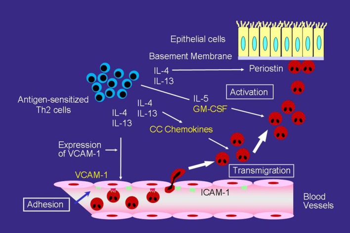
Figure 1
Full legend
Mechanisms of eosinophilic airway inflammation in bronchial asthma. IL-5 plays an important role in the development of eosinophilic airway inflammation. However, in the absence of IL-5, the Th2 network, which includes a cascade of VCAM-1/CC chemokines/GM-CSF, can maintain eosinophilic infiltration and activation. Yellow text indicates a cascade of VCAM-1/CC chemokines/GM-CSF.
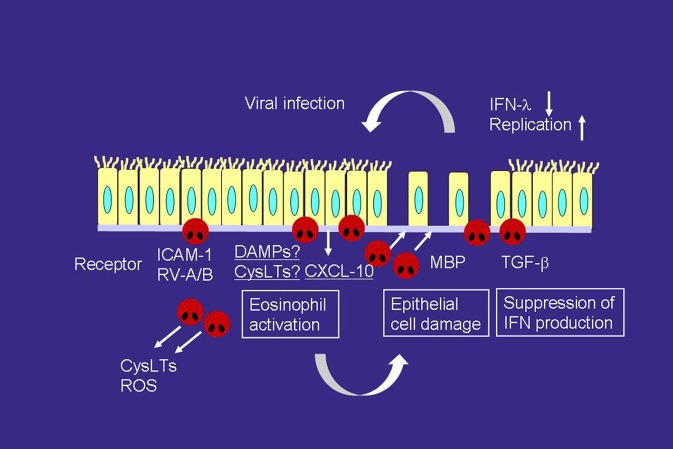
Figure 2
Full legend
Interactions of viral infection and eosinophils in the development of asthma exacerbation. RV infection releases a variety of mediators, including CXCL-10 and cysLTs, which can directly activate eosinophils and induce asthma exacerbation, from airway epithelial cells. RV infection increases the expression of ICAM-1, which also activates eosinophils. On the other hand, activated eosinophils release MBP, which induces epithelial cell damage. Eosinophils also produce TGF-β, which can suppress the expression of IFN-λ, anti-viral cytokines, on epithelial cells. Therefore, eosinophilic airway inflammation can increase susceptibility to RV infection. ROS, reactive oxygen species; TGF, transforming growth factor.
References107
- 1.
Bochner BS Undem BJ Lichtenstein LM. Immunological aspects of allergic asthma. Annu Rev Immunol. (1994 ) 12:295–335.
Not in our library10.1146/annurev.iy.12.040194.001455 (opens in a new tab)
- 2.
Barnes PJ. Pathophysiology of allergic inflammation. Immunol Rev. (2011 ) 242:31–50.
Not in our library10.1111/j.1600-065x.2011.01020.x (opens in a new tab)
- 3.
Lötvall J Akdis CA Bacharier LB Bjermer L Casale TB Custovic A. Asthma endotypes: a new approach to classification of disease entities within the asthma syndrome. J Allergy Clin Immunol. (2011 ) 127:355–60.
Not in our library10.1016/j.jaci.2010.11.037 (opens in a new tab)
- 4.
Wenzel SE. Asthma phenotypes: the evolution from clinical to molecular approaches. Nat Med. (2012 ) 18:716–25.
Not in our library10.1038/nm.2678 (opens in a new tab)
- 5.
Green RH Brightling CE McKenna S Hargadon B Parker D Bradding P. Asthma exacerbations and sputum eosinophil counts: a randomised controlled trial. Lancet (2002 ) 360:1715–21.
Not in our library10.1016/s0140-6736(02)11679-5 (opens in a new tab)
- 6.
Price DB Rigazio A Campbell JD Bleecker ER Corrigan CJ Thomas M. Blood eosinophil count and prospective annual asthma disease burden: a UK cohort study. Lancet Respir Med. (2015 ) 3, 849–58.
Not in our library10.1016/s2213-2600(15)00367-7 (opens in a new tab)
- 7.
Denlinger LC Phillips BR Ramratnam S Ross K Bhakta NR Cardet JC. Inflammatory and comorbid features of patients with severe asthma and frequent exacerbations. Am J Respir Crit Care Med. (2017 ) 195:302–13.
Not in our library10.1164/rccm.201602-0419oc (opens in a new tab)
- 8.
Weller PF Human eosinophils. J Allergy Clin Immunol. (1997 ) 100:283–7.
Not in our library10.1016/s0091-6749(97)70237-9 (opens in a new tab)
- 9.
Gleich GJ. Mechanisms of eosinophil-associated inflammation. J Allergy Clin Immunol. (2000 ) 105:651–63.
Not in our library10.1067/mai.2000.105712 (opens in a new tab)
- 10.
Leckie MJ ten Brinke A Khan J Diamant Z O'Connor BJ Walls CM (2000 ). Effects of an interleukin-5 blocking monoclonal antibody on eosinophils, airway hyper-responsiveness, and the late asthmatic response. Lancet 356, 2144–2148.
Not in our library10.1016/s0140-6736(00)03496-6 (opens in a new tab)
- 11.
Flood-Page P Swenson C Faiferman I Matthews J Williams M Brannick L. A study to evaluate safety and efficacy of mepolizumab in patients with moderate persistent asthma. Am J Respir Crit Care Med. (2007 ) 176:1062–71.
Not in our library10.1164/rccm.200701-085oc (opens in a new tab)
- 12.
Kay AB Phipps S Robinson DS. A role for eosinophils in airway remodelling in asthma. Trends Immunol. (2004 ) 25:477–82.
Not in our library10.1016/j.it.2004.07.006 (opens in a new tab)
- 13.
Humbles AA Lloyd CM McMillan SJ Friend DS Xanthou G McKenna EE. A critical role for eosinophils in allergic airways remodeling. Science (2004 ) 305:1776–9.
Not in our library10.1126/science.1100283 (opens in a new tab)
- 14.
Wong DT Elovic A Matossian K Nagura N McBride J Chou MY Eosinophils from patients with blood eosinophilia express transforming growth factor β1. Blood (1991 ) 78:2702–7.
Not in our library
- 15.
Owen WF Jr Soberman RJ Yoshimoto T Sheffer AL Lewis RA Austen KF. Synthesis and release of leukotriene C4 by human eosinophils. J Immunol. (1987 ) 138:532–8.
Not in our library
- 16.
Cowburn AS Sladek K Soja J Adamek L Nizankowska E Szczeklik A. Overexpression of leukotriene C4 synthase in bronchial biopsies from patients with aspirin-intolerant asthma. J Clin Invest. (1998 ) 101:834–46.
Not in our library10.1172/jci620 (opens in a new tab)
- 17.
Seymour ML Rak S Aberg D Riise GC Penrose JF Kanaoka Y. Leukotriene and prostanoid pathway enzymes in bronchial biopsies of seasonal allergic asthmatics. Am J Respir Crit Care Med. (2001 ) 164:2051–6.
Not in our library10.1164/ajrccm.164.11.2008137 (opens in a new tab)
- 18.
Flood-Page P Menzies-Gow A Phipps S Ying S Wangoo A Ludwig MS. Anti-IL-5 treatment reduces deposition of ECM proteins in the bronchial subepithelial basement membrane of mild atopic asthmatics. J Clin Invest. (2003 ) 112, 1029–1036.
Not in our library10.1172/jci17974 (opens in a new tab)
- 19.
Nair P Pizzichini MM Kjarsgaard M Inman MD Efthimiadis A Pizzichini E. Mepolizumab for prednisone-dependent asthma with sputum eosinophilia. N Engl J Med. (2009 ) 360:985–93.
Not in our library10.1056/nejmoa0805435 (opens in a new tab)
- 20.
Haldar P Brightling CE Hargadon B Gupta S Monteiro W Sousa A. Mepolizumab and exacerbations of refractory eosinophilic asthma. N Engl J Med. (2009 ) 360:973–84.
Not in our library10.1056/nejmoa0808991 (opens in a new tab)
- 21.
Pavord ID Korn S Howarth P Bleecker ER Buhl R Keene ON. Mepolizumab for severe eosinophilic asthma (DREAM): a multicentre, double-blind, placebo-controlled trial. Lancet (2012 ) 380:651–9.
Not in our library10.1016/s0140-6736(12)60988-x (opens in a new tab)
- 22.
Gleich GJ Flavahan NA Fujisawa T Vanhoutte PM. The eosinophil as a mediator of damage to respiratory epithelium: a model for bronchial hyperreactivity. J Allergy Clin Immunol. (1988 ) 81:776–81.
Not in our library10.1016/0091-6749(88)90931-1 (opens in a new tab)
- 23.
Wardlaw AJ Dunnette S Gleich GJ Collins JV Kay AB. Eosinophils and mast cells in bronchoalveolar lavage in subjects with mild asthma. Relation. bronchial hyperreactivity. Am Rev Respir Dis. (1988 ) 137:62–9.
Not in our library10.1164/ajrccm/137.1.62 (opens in a new tab)
- 24.
Kelly EA Esnault S Liu LY Evans MD Johansson MW Mathur S Mepolizumab attenuates airway eosinophil numbers, but not their functional phenotype, in asthma. Am J Respir Crit Care Med. (2017 ) 196:1385–95.
Not in our library10.1164/rccm.201611-2234oc (opens in a new tab)
- 25.
Nakagome K Nagata M. Pathogenesis of airway inflammation in bronchial asthma. Auris Nasus Larynx (2011 ) 38:555–63.
Not in our library10.1016/j.anl.2011.01.011 (opens in a new tab)
- 26.
Schleimer RP Sterbinsky SA Kaiser J Bickel CA Klunk DA Tomioka K (1992 ). IL-4 induces adherence of human eosinophils and basophils but not neutrophils to endothelium. Association with expression of VCAM-1. J Immunol. 148:1086–92.
Not in our library
- 27.
Bochner BS. Cellular adhesion and its antagonism. J Allergy Clin Immunol. (1997 ) 100:581–5.
Not in our library10.1016/s0091-6749(97)70158-1 (opens in a new tab)
- 28.
Nagata M Sedgwick JB Bates ME Kita H Busse WW. Eosinophil adhesion to vascular cell adhesion molecule-1 activates superoxide anion generation. J Immunol. (1995 ) 155:2194–202.
Not in our library
- 29.
Nagata M Sedgwick JB Kita H Busse WW. Granulocyte macrophage colony-stimulating factor augments ICAM-1 and VCAM-1 activation of eosinophil function. Am J Respir Cell Mol Biol. (1998 ) 19:158–66.
Not in our library10.1165/ajrcmb.19.1.3001 (opens in a new tab)
- 30.
Nagata M Sedgwick JB Vrtis R Busse WW. Endothelial cells upregulate eosinophil superoxide generation via VCAM-1 expression. Clin Exp Allergy (1999 ) 29:550–61.
Not in our library10.1046/j.1365-2222.1999.00506.x (opens in a new tab)
- 31.
Nagata M Yamamoto H Tabe K Sakamoto Y. Eosinophil transmigration across VCAM-1-expressing endothelial cells is upregulated by antigen-stimulated mononuclear cells. Int Arch Allergy Immunol. (2001 ) 125:7–11.
Not in our library10.1159/000053844 (opens in a new tab)
- 32.
Ying S Robinson DS Meng Q Rottman J Kennedy R Ringler DJ. Enhanced expression of eotaxin and CCR3 mRNA and protein in atopic asthma. Association with airway hyperresponsiveness and predominant co-localization of eotaxin mRNA to bronchial epithelial and endothelial cells Eur J Immunol. (1997 ) 27:3507–16.
Not in our library10.1002/eji.1830271252 (opens in a new tab)
- 33.
Nakagome K Shoda H Shirai T Nishihara F Soma T Uchida Y. Eosinophil transendothelial migration induced by the bronchoalveolar lavage fluid of acute eosinophilic pneumonia. Respirology (2017 ) 22:913–21.
Not in our library10.1111/resp.12982 (opens in a new tab)
- 34.
Liu LY Sedgwick JB Bates ME Vrtis RF Gern JE Kita H. (2002 ). Decreased expression of membrane IL-5 receptor alpha on human eosinophils: I. Loss of membrane IL-5 receptor alpha on airway eosinophils and increased soluble IL-5 receptor alpha in the airway after allergen challenge. J Immunol. 169:6452–8.
Not in our library10.4049/jimmunol.169.11.6452 (opens in a new tab)
- 35.
Liu LY Sedgwick JB Bates ME Vrtis RF Gern JE Kita H. (2002 ). Decreased expression of membrane IL-5 receptor alpha on human eosinophils: II. IL-5 down-modulates its receptor via a proteinase-mediated process. J Immunol. 169:6459–66.
Not in our library10.4049/jimmunol.169.11.6459 (opens in a new tab)
- 36.
Laitinen LA Laitinen A Haahtela T Vilkka V Spur BW Lee TH Leukotriene E4 and granulocytes infiltration into asthmatic airways. Lancet (1993 ) 341:989–90.
Not in our library10.1016/0140-6736(93)91073-u (opens in a new tab)
- 37.
Nagata M Saito K Tsuchiya T Sakamoto Y. Leukotriene D4 upregulates eosinophil adhesion via the cysteinyl leukotriene 1 receptor. J Allergy Clin Immunol. (2002 ) 109:676–80.
Not in our library10.1067/mai.2002.122841 (opens in a new tab)
- 38.
Saito K Nagata M Kikuchi I Sakamoto Y. Leukotriene D4 and eosinophil transendothelial migration, superoxide generation, and degranulation via β2 integrin. Ann Allergy Asthma Immunol. (2004 ) 93:594–600.
Not in our library10.1016/s1081-1206(10)61269-0 (opens in a new tab)
- 39.
Pizzichini E Leff JA Reiss TF Hendeles L Boulet LP Wei LX. Montelukast reduces airway eosinophilic inflammation in asthma: a randomized, controlled trial. Eur Respir J. (1999 ) 14:12–8.
Not in our library10.1034/j.1399-3003.1999.14a04.x (opens in a new tab)
- 40.
Obase Y Shimoda T Tomari SY Mitsuta K Kawano T Matsuse H. Effects of pranlukast on chemical mediators in induced sputum on provocation tests in atopic and aspirin-intolerant asthmatic patients. Chest (2002 ) 121:143–50.
Not in our library10.1378/chest.121.1.143 (opens in a new tab)
Cited by papers we hold1
Reads
Read by people on this page
None yet.
Checked or read by AI through naulon
None yet.
A person counts once a day per paper. We keep no cookie and no address: a reader is a hash that changes every day. Any month under five reads shows no number.
Are you an author?
Is this your paper?
Claim it to add your note and be paid when agents cite it. Free.