Isolation and Characterization of CD34+ Blast-Derived Exosomes in Acute Myeloid Leukemia
Chang Sook Hong, Laurent Muller, Michael Boyiadzis and 1 more
PLoS ONE · 2014
doi:10.1371/journal.pone.0103310 (opens in a new tab)
We email you when a new notice or a question about this paper appears.
Abstract
Exosomes are membrane-bound vesicles found in all biological fluids. AML patients' plasma collected at diagnosis contains elevated exosome levels relative to normal donor (ND) plasma. The molecular profile of AML exosomes changes in the course of therapy and may serve as a measure of disease progression or response to therapy. However, plasma contains a mix of exosomes derived from various cell types. To be able to utilize blast-derived exosomes as biomarkers for AML, we have developed an immunoaffinity-based capture method utilizing magnetic microbeads coated with anti-CD34 antibody (Ab). This Ab is specific for CD34, a unique marker of AML blasts. The capture procedure was developed using CD34+ exosomes derived from Kasumi-1 AML cell culture supernatants. The capture capacity of CD34microbeads was shown to linearly correlate with the input exosomes. A 10 uL aliquot of CD34 microbeads was able to capture all of CD34+ exosomes present in 100-1,000 uL of AML plasma. The levels of immunocaptured CD34+ exosomes correlated with the percentages of CD34+ blasts in the AML patients' peripheral blood. The immunocaptured exosomes had a typical cup-shaped morphology by transmission electron microscopy, and their molecular cargo was similar to that of parental blasts. These exosomes were biologically-active. Upon co-incubation with natural killer (NK) cells, captured blast-derived exosomes down-regulated surface NKG2D expression, while non-captured exosomes reduced expression levels of NKp46. Our data provide a proof-of-principle that blast-derived exosomes can be quantitatively recovered from AML patients' plasma, their molecular profile recapitulates that of autologous blasts and they retain the ability to mediate immune suppression. These data suggest that immunocaptured blast-derived exosomes might be useful in diagnosis and/or prognosis of AML in the future.
Figures6
From PubMed Central, U.S. National Library of Medicine.
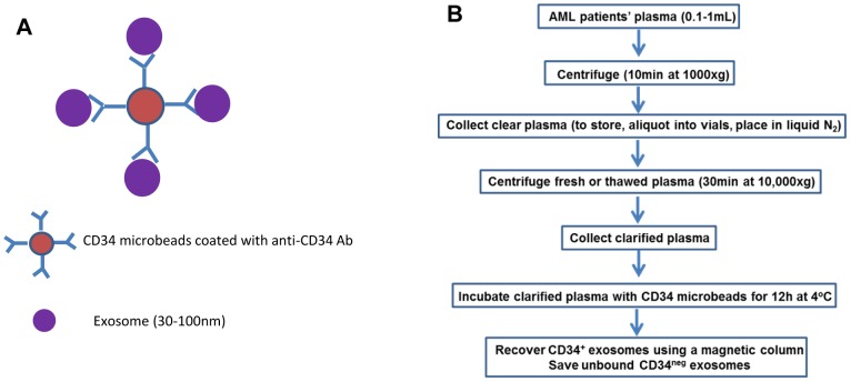
Figure 1
Full legend
The strategy for capture of CD34+ exosomes using anti-CD34 Ab-coated microbeads purchased from Miltenyi (A) and a schema for capture of CD34+ exosomes directly from AML patients' plasma (B).
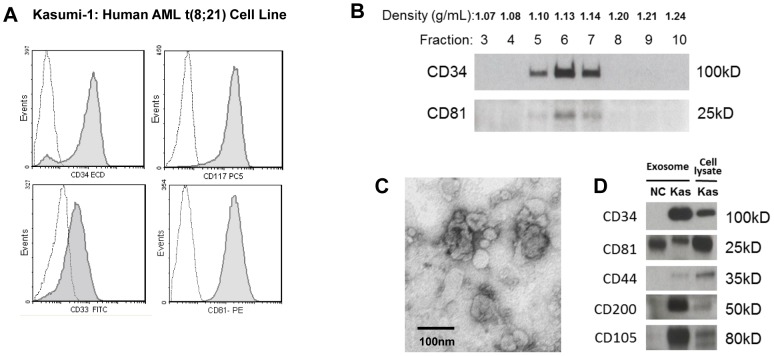
Figure 2
Full legend
Studies with the Kasumi-1 cell line and with exosomes derived from Kasumi-1 culture supernatants. A) Flow cytometry of Kasumi-1 cells shows expression of blast markers, CD34, CD117 and CD33 and of CD81 (tetraspanin). Dotted lines indicate isotype controls. B) Kasumi-1 exosomes were separated on a continuous sucrose density gradient, and the collected gradient fractions were tested in Western blots for CD34 and CD81. CD34+ and CD81+ exosomes were recovered at the density of 1.10–1.14. C) An electron microscope image of isolated and negatively stained Kasumi-1 exosomes. D) Western blots of isolated Kasumi-1 exosomes (Kas) and of exosomes isolated from plasma of a normal donor (NC). Each lane was loaded with 10 µg exosomal protein.
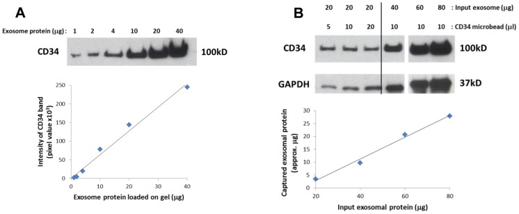
Figure 3
Full legend
Calibration experiments with CD34+ Kasumi-1 exosomes isolated by ultracentrifugation. A) Isolated exosomes were loaded at increasing protein concentrations onto gels and blotted using anti-CD34 Ab. Intensity of each band was measured in pixels. The graph illustrates a linear relationship between exosomal protein levels and pixel intensity in Western blots. B) Isolated exosomes (20 µg protein) were added to increasing volumes of CD34 microbeads (left). Five or 10 µL of microbeads were sufficient to capture 20 µg of exosomes. Next, a 10 µL aliquot of beads was used to capture increasing concentrations of Kasumi-1 exosomes (right). The graph shows that the capacity of CD34 microbeads to capture up to 80 µg of input exosomes increased linearly. However, at 80 µg only about 40% of exosomal proteins were captured, suggesting that additional microbeads are necessary for capture of all Kasumi-1 exosomes.
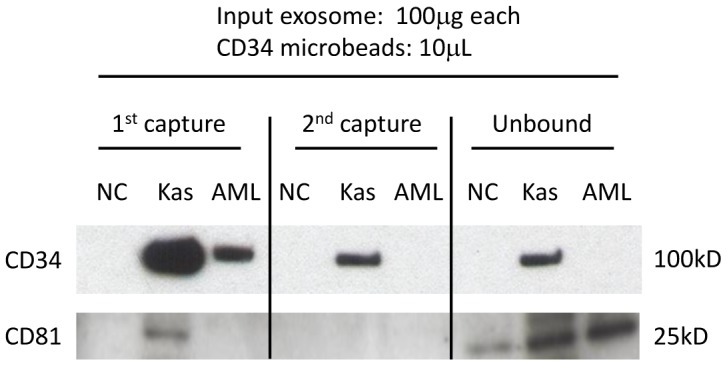
Figure 4
Full legend
Capture of CD34+ exosomes from total exosomal fractions by CD34+ microbeads. Isolated Kasumi-1 exosomes (Kas) and total exosome fractions isolated by ultracentrifugation from normal donor plasma (NC) or from CD34+ AML patient plasma (AML) were used for capture with CD34 microbeads. After the 1st capture and removal of beads, 2nd capture was performed with a fresh aliquot of CD34 microbeads. The final unbound fractions were ultracentrifuged to collect remaining exosomes. While the 2nd capture was necessary to recover all CD34+ Kasumi-1 exosomes, a single capture was sufficient to recover all CD34+ exosomes from the bulk of exosomes isolated from the AML plasma. There were no CD34+ exosomes captured from the bulk exosomal fraction isolated from a normal donor's plasma. CD81 expression indicates that the unbound fractions still contain CD34neg exosomes.
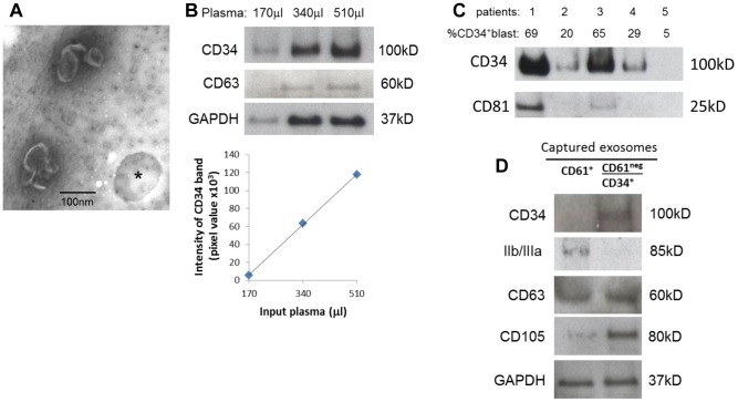
Figure 5
Full legend
Capture of CD34+ blast-derived exosomes directly from AML patients' plasma. A) Negatively stained electron microscope images of exosomes captured on CD34 microbeads (* shows a vacant microbead). B) Increasing AML plasma volumes were used for capture of CD34+ exosomes by microbeads and recovered exosomes were studied by Western blots. The graph shows a linear relationship between the input plasma volumes and pixel densities of captured and blotted CD34+ exosomes. C) Exosomes were captured from plasma samples obtained from five CD34+ AML patients and were analyzed by Western blots. The percentage of leukemic blasts in the peripheral circulation of each of the patients is shown. CD81 serves as the exosome marker. D) Removal of platelet-derived exosomes from plasma using anti-CD61 Ab-coated microbeads prior to capture of CD34+ exosomes. Exosomes captured with CD61 microbeads (left: CD61+) and CD34+ exosomes captured after removing CD61+ exosomes (right: CD61neg/CD34+) are shown in a representative Western blot of three evaluated.
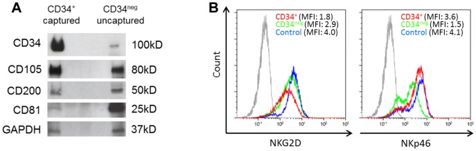
Figure 6
Full legend
Characteristics of captured exosomes. A) A representative Western blot profile of the captured CD34+ exosomes and of non-captured CD34neg exosomes which were isolated by ultracentrifugation from the same AML plasma sample. B) After co-incubation of NK cells with CD34+ exosomes captured directly from AML plasma or CD34neg exosomes as described in materials and Methods, NKG2D expression (in MFI) was found to be down-regulated only with CD34+ exosomes (red) compared to CD34neg exosomes (green) or control without exosomes (blue). The gray peak denotes isotype control. In contrast, NKp46 expression was down-regulated by co-incubation with CD34neg exosomes.
References27
- 1.
Burnett A, Wetzler M, Lowenberg B (2011 ) Therapeutic advances in acute myeloid leukemia. J Clin Onc 29(5): 487–94.
Not in our library10.1200/jco.2010.30.1820 (opens in a new tab)
- 2.
Horton SJ, Huntly BJ (2012 ) Recent advances in acute myeloid leukemia stem cell biology. Haematologica 97: 966–74.
Not in our library10.3324/haematol.2011.054734 (opens in a new tab)
- 3.
EL Andaloussi S, Mäger I, Breakefield XO, Wood MJ (2013 ) Extracellular vesicles: biology and emerging therapeutic opportunities. Nat Rev Drug Discov 12: 347–57.
Not in our library10.1038/nrd3978 (opens in a new tab)
- 4.
Peinado H, Aleckovic M, Lavotshkin S, Matei I, Costa-Silva B, et al (2012 ) Melanoma exosomes educate bone marrow progenitor cells toward a pro-metastatic phenotype through MET. Nat Med 18: 883–91.
Not in our library10.1038/nm.2753 (opens in a new tab)
- 5.
Filipazzi P, Burdek M, Villa A, Rivoltini L, Huber V (2012 ) Recent advances on the role of tumor exosomes in immunosuppression and disease progression. Semin Cancer Biol 22: 342–9.
Not in our library10.1016/j.semcancer.2012.02.005 (opens in a new tab)
- 6.
Whiteside TL (2013 ) Immune modulation of T-cell and NK (natural killer) cell activities by TEXs (tumour-derived exosomes). Biochem Soc Transact 41: 245–51.
Not in our library10.1042/bst20120265 (opens in a new tab)
- 7.
Thery C, Zitvogel L, Amigorena S (2002 ) Exosomes: composition, biogenesis and function. Nat Rev Immunol 2: 569–79.
Not in our library10.1038/nri855 (opens in a new tab)
- 8.
Zhang HG, Zhuang X, Sun D, Liu Y, Xiang X, et al (2012 ) Exosomes and immune surveillance of neoplastic lesions: a review. Biotech Histochem 87: 161–8.
Not in our library10.3109/10520291003659042 (opens in a new tab)
- 9.
Taylor DD, Gercel-Taylor C (2011 ) Exosomes/microvesicles: mediators of cancer-associated immunosuppressive microenvironments. Semin Immunopathol 33: 441–54.
Not in our library10.1007/s00281-010-0234-8 (opens in a new tab)
- 10.
Martínez-Lorenzo MJ, Anel A, Alava MA, Piñeiro A, Naval J, et al (2004 ) The human melanoma cell line MelJuSo secretes bioactive FasL and APO2L/TRAIL on the surface of microvesicles. Possible contribution to tumor counterattack. Exp Cell Res 295: 315–29.
Not in our library10.1016/j.yexcr.2003.12.024 (opens in a new tab)
- 11.
Andreola G, Rivoltini L, Castelli C, Huber V, Perego P, et al (2002 ) Induction of lymphocyte apoptosis by tumor cell secretion of FasL-bearing microvesicles. J Exp Med 195: 1303–16.
Not in our library10.1084/jem.20011624 (opens in a new tab)
- 12.
Valenti R, Huber V, Filipazzi P, Pilla L, Sovena G, et al (2006 ) Human tumor-released microvesicles promote the differentiation of myeloid cells with transforming growth factor-beta-mediated suppressive activity on T lymphocytes. Cancer Res 66: 9290–8.
Not in our library10.1158/0008-5472.can-06-1819 (opens in a new tab)
- 13.
Szczepanski MJ, Szajnik M, Welsh A, Whiteside TL, Boyiadzis M (2011 ) Blast-derived microvesicles in sera from patients with acute myeloid leukemia suppress natural killer cell function via membrane-associated transforming growth factor-beta1. Haematologica 96: 1302–9.
Not in our library10.3324/haematol.2010.039743 (opens in a new tab)
- 14.
- 15.
Muller L, Hong C-S, Stolz DR, Watkins S, Whiteside TL (2014) Isolation of biologically active exosomes from human plasma. Ms. submitted.
Not in our library10.1016/j.jim.2014.06.007 (opens in a new tab)
- 16.
Aatonen M, Grönholm M, Siljander PR (2012 ) Platelet-derived microvesicles: multitalented participants in intercellular communication. Semin Thromb Hemost 38: 102–13.
Not in our library10.1055/s-0031-1300956 (opens in a new tab)
- 17.
Asou H, Tashiro S, Hamamoto K, Otsuji A, Kita K, et al (1991 ) Establishment of a human acute myeloid leukemia cell line (Kasumi-1) with 8;21 chromosome translocation, Blood. 77: 2031–2036.
Not in our library
- 18.
Larizza L, Magnani I, Beghini A (2005 ) The Kasumi-1 cell line: a t(8;21)-kit mutant model for acute myeloid leukemia. Leuk Lymphoma 46: 247–55.
Not in our library10.1080/10428190400007565 (opens in a new tab)
- 19.
Teague RM, Kline J (2013 ) Immune evasion in acute myeloid leukemia: current concepts and future directions. J Immunother Cancer 1 13.
Not in our library10.1186/2051-1426-1-13 (opens in a new tab)
- 20.
Whiteside TL (2014 ) Regulatory T cell subsets in human cancer: are they regulating for or against tumor progression? Cancer Immunol Immunother 63: 67–72.
Not in our library10.1007/s00262-013-1490-y (opens in a new tab)
- 21.
- 22.
Mathivanan S, Lim JW, Tauro BJ, Ji H, Moritz RL, et al (2010 ) Proteomics analysis of A33 immunoaffinity-purified exosomes released from the human colon tumor cell line LIM1215 reveals a tissue-specific protein signature. Mol Cell Proteomics 9: 197–208.
Not in our library10.1074/mcp.m900152-mcp200 (opens in a new tab)
- 23.
Tauro BJ, Greening DW, Mathias RA, Mathivanan S, Ji H, et al (2013 ) Two distinct populations of exosomes are released from LIM1863 colon carcinoma cell-derived organoids. Mol Cell Proteomics 12: 587–98.
Not in our library10.1074/mcp.m112.021303 (opens in a new tab)
- 24.
van Stijn A, van der Pol MA, Kok A, Bontje PM, et al (2003 ) Differences between the CD34+ and CD34- blast compartments in apoptosis resistance in acute myeloid leukemia. Haematologica 88: 497–508.
Not in our library
- 25.
Ishikawa F1, Yoshida S, Saito Y, Hijikata A, Kitamura H, et al (2007 ) Chemotherapy-resistant human AML stem cells home to and engraft within the bone-marrow endosteal region. Nat Biotechnol 25: 1315–21.
Not in our library10.1038/nbt1350 (opens in a new tab)
- 26.
Oyan AM, Bø TH, Jonassen I, Ulvestad E, Gjertsen BT, et al (2005 ) CD34 expression in native human acute myelogenous leukemia blasts: differences in CD34 membrane molecule expression are associated with different gene expression profiles. Cytometry B Clin Cytom 64: 18–27.
Not in our library10.1002/cyto.b.20044 (opens in a new tab)
- 27.
Blair A, Hogge DE, Sutherland HJ (1998 ) Most acute myeloid leukemia progenitor cells with long-term proliferative ability in vitro and in vivo have the phenotype CD34+/CD71-/HLA-DR+. Blood 92: 4325–4335.
Not in our library
Cited by papers we hold15
Reads
Read by people on this page
None yet.
Checked or read by AI through naulon
None yet.
A person counts once a day per paper. We keep no cookie and no address: a reader is a hash that changes every day. Any month under five reads shows no number.
Are you an author?
Is this your paper?
Claim it to add your note and be paid when agents cite it. Free.