Factors associated with disease severity and mortality among patients with COVID-19: A systematic review and meta-analysis
Vignesh Chidambaram, Nyan Lynn Tun, Waqas Z. Haque and 23 more
PLOS ONE · 2020 · Meta-analysis · CC BY
doi:10.1101/2020.08.07.20166868 (opens in a new tab)
We email you when a new notice or a question about this paper appears.
Abstract
Background Understanding the factors associated with disease severity and mortality in Coronavirus disease (COVID-19) is imperative to effectively triage patients. We performed a systematic review to determine the demographic, clinical, laboratory and radiological factors associated with severity and mortality in COVID-19. Methods We searched PubMed, Embase and WHO database for English language articles from inception until May 8, 2020. We included Observational studies with direct comparison of clinical characteristics between a) patients who died and those who survived or b) patients with severe disease and those without severe disease. Data extraction and quality assessment were performed by two authors independently. Results Among 15680 articles from the literature search, 109 articles were included in the analysis. The risk of mortality was higher in patients with increasing age, male gender (RR 1.45, 95%CI 1.23–1.71), dyspnea (RR 2.55, 95%CI 1.88–2.46), diabetes (RR 1.59, 95%CI 1.41–1.78), hypertension (RR 1.90, 95%CI 1.69–2.15). Congestive heart failure (OR 4.76, 95%CI 1.34–16.97), hilar lymphadenopathy (OR 8.34, 95%CI 2.57–27.08), bilateral lung involvement (OR 4.86, 95%CI 3.19–7.39) and reticular pattern (OR 5.54, 95%CI 1.24–24.67) were associated with severe disease. Clinically relevant cut-offs for leukocytosis(>10.0 x10 9 /L), lymphopenia(< 1.1 x10 9 /L), elevated C-reactive protein(>100mg/L), LDH(>250U/L) and D-dimer(>1mg/L) had higher odds of severe disease and greater risk of mortality. Conclusion Knowledge of the factors associated of disease severity and mortality identified in our study may assist in clinical decision-making and critical-care resource allocation for patients with COVID-19.
Full text
From PubMed Central, U.S. National Library of Medicine, as current on Oct 24, 2026. Paragraph numbers are ours, so an answer can point at one.
Introduction
Since the first documented reports of the severe acute respiratory syndrome coronavirus 2 (SARS-CoV-2) infection, the virus has had a global impact, affecting millions, which led the World Health Organization (WHO) to declare the outbreak a pandemic. Patients who develop Coronavirus Disease 2019 (COVID-19) may require hospitalization and intensive care unit admission.
With variable access to critical care resources across countries, recent guidelines for the COVID-19 pandemic have called for allocating life sustaining treatments based on a patient’s risk of mortality. Health systems preparedness requires a deeper understanding of how to effectively triage patients with COVID-19, in order to maximize the benefit of scarce intensive care unit resources while minimizing the potential harm of outpatient management of ill patients. While the guidelines warrant utilizing triage scores that have been previously validated for assessing organ failure/dysfunction and survival (e.g. sequential organ failure assessment, SOFA), these are non-specific in etiology. Thus, having an understanding of the predisposing conditions and disease-specific clinical, laboratory and radiological parameters, may lay the groundwork for developing a COVID-19 specific composite score at a later stage, which can predict unfavorable clinical outcomes.
With the massive influx of studies on COVID-19 in the recent months and their often conflicting or unclear findings, a systematic review of the factors associated with survival or disease severity in patients with COVID-19 that takes into consideration the inherent variability in study population, will be of great utility to clinicians, researchers and policy makers. In this systematic review and meta-analysis, we sought to better understand the clinical, laboratory and radiological parameters associated with mortality and disease severity among patients with COVID-19.
Search strategy and study selection
We followed the PRISMA guidelines for reporting in systematic reviews and meta-analyses. We searched PubMed, Embase and the WHO COVID-19 database by using the search strategy included in the supplementary document in S1 Appendix (Section I). For PubMed and Embase, an initial search on March 26, 2020 was conducted, and updated multiple times with the final update performed on May 7, 2020. The WHO database was initially downloaded on March 27, 2020 and the final update was performed on May 8, 2020. Only articles published in the English language were included. We only included article published in peer-reviewed academic journals; we did not include articles uploaded in the preprint servers, as they are not peer reviewed and the findings may not be reliable.
We included observational studies that included patients with microbiologically confirmed SARS-CoV-2 infection, irrespective of the age of the participants. The study designs of the included studies were assessed and recorded independently by two authors (VC and MM) acting as arbiters. The differentiation between case series and cohort studies was made based on the criteria outlined by Dekkers et al. Case reports, case series, and randomized control trials were excluded from this review. We included all studies that reported a direct comparison of clinical, laboratory or radiologic characteristics between a) patients who died and those who survived or b) patients with severe disease and those without severe disease. Only those studies which defined “severe disease” based on the American Thoracic Society guidelines for the treatment of Community-acquired Pneumonia or the Chinese National Health Commission guidelines for the Treatment of Novel Coronavirus infection, were included in our analysis. Studies which only described the characteristics of patients who died or patients with severe disease were excluded if there was no comparison group.
We only included studies reporting primary hospital data on patients, while studies with centralized data from national health agencies and databases were excluded from our review. Efforts were made, as feasible, to minimize overlap of patients across studies by collecting information on the name of the hospital, date of hospital admission of the participants and the names of the investigators.
Literature screening
The COVIDENCE platform was used for conducting this systematic review. After de-duplication, the titles and abstracts of the articles retrieved from the search strategy were screened independently by at least two of the following authors (VC, NT, WH, RS, AK, AH, II, AN, SA, ES, and MS) and conflicts were resolved by consensus between VC and NT. The full texts of the articles included after title and abstract screening were independently screened by at least two of the following authors (VC, NT, WH, RS, AK, AH, II, AN, SA, ES, and MS) and conflicts were resolved based on consensus between VC and NT. Specific reasons for study exclusion are listed in Fig 1.
Data extraction and quality assessment
Data extraction was performed independently by at least two of the authors (VC, NT, WH, MM, RS, AK, AH, II, AN, SA, ES, MS, AS, KW, and TP) and conflicts were resolved by a consensus between two authors (MM and VC). The data extraction form for this review was created using the Qualtrics platform. The primary outcomes were a) death of the patient and b) the presence of severe disease in the patient. There were no secondary outcomes. Data on the study characteristics, source of funding, demographic characteristics, comorbidities, clinical symptoms, in-hospital complications, laboratory, and radiological features of the study participants were extracted. Data on sex of the patient, smoking status, presence of comorbidities, clinical symptoms, in-hospital complications, and radiological features were extracted as binary variables, while age was extracted as continuous data. For the laboratory parameters, data were extracted as both continuous and categorical variables. Binary data for the laboratory variables were collected using all the cut-offs described in the included studies. The time points for lab measurements, radiological evaluation and the disease severity assessment were extracted for the included studies. Continuous data for the age and laboratory variables were documented as mean. The median values reported in studies were transformed into mean.
The risk assessment for bias for all the studies included in this review was performed using the Newcastle-Ottawa quality assessment scale (NOS) for observational and cohort studies. The three major domains of quality of a study covered by this tool were selection of participants, comparability of cohorts and outcome assessment against a total score of 9. This was performed independently by at least two of the following authors (RS, MM, ES, SA). When conflicts related to bias arose, the final decision was taken by a consensus between VC and NT.
Data synthesis and analysis
We performed a meta-analysis with random effects model to obtain pooled effect sizes for the outcomes of interest. The associations between binary parameters and mortality were reported using pooled risk ratios (with 95%CI). Due to the lack of consistency in the time point of assessment of disease severity, odds ratios (with 95%CI) were used to determine the association between the various factors and the presence of severe disease. When the laboratory parameters were reported using different cut-offs, we reported the effect sizes for each cut-off taken separately in addition to the pooled effect sizes for all the cut-offs taken together. Statistical heterogeneity across the studies was assessed by forest plots, I2 and Tau2 statistics. When the I2 was more than 60%, we performed subgroup analyses based on the whether the studies included all patients with COVID-19 or only the patients who were critically ill. If the heterogeneity was still higher than 60%, we performed sensitivity analyses by excluding studies with a low quality (NOS ≤ 5). Sensitivity analyses for the laboratory and radiological parameters were also performed by excluding studies which did not report the time points of assessment. Publication bias was assessed by visual inspection of the funnel plot, and Egger’s test was performed for those exposures reported by at least 10 studies. For continuous variables, meta-regression was performed to assess the percentage change in mortality or the presence of severe disease with unit increase in the mean of laboratory parameter reported in the studies. For the binary exposures, unadjusted effect sizes were calculated from the summary data. Adjusted effect sizes were not used due to the lack of consistency in the parameters that they are adjusted for, among the included studies. All analyses were carried out using the meta package in Stata (StataCorp, version 16).
Role of the funding source
This study was not supported by any funding source.
Results
We identified 15680 studies from three databases after removing duplicates, of which the full text was retrieved for 502 articles. All the articles included were in the English language. The reasons for exclusion of studies are outlined in Fig 1. A total of 109 studies were included for this review, of which 42 studies assessed mortality risks; 72 studies determined association with severe disease, out of which 5 studies reported both the outcomes. Of the total 109 studies in the review, 101 were retrospective cohorts, seven were prospective cohorts, and one of them was an ambispective cohort study. There were no cross-sectional or case control studies that satisfied the inclusion criteria. Of the studies that determined the risk of mortality, an aggregate of 20296 participants were assessed, with 32 studies from China, six from the United States, two from Spain, one from the United Kingdom, one from Italy, one from Iran and a multi-country study. Five of these studies only included patients who were critically ill or invasively ventilated. Among the studies that assessed the association with severe disease, a total of 17992 participants were included with seventy-one studies from China and one study from Italy. The characteristics of the included studies, along with the time points for laboratory, radiological and disease severity assessment, are outlined in the S1 and S2 Tables (in S1 Appendix Section II).
Quality assessment was performed using the New-Castle Ottawa scale (NOS) as all of the studies used a cohort study design. This revealed that one of the studies (0.9%) had scored 9, 54 studies (49.5%) had scored 8, 39 studies (35.7%) scored 7, six studies (5.5%) scored 6. A total of nine studies were identified as low-quality studies (NOS ≤ 5) with six studies (5.5%) scoring 5, and the remaining three studies (2.7%) scoring 4 (S3 and S4 Tables—in S1 Appendix section III).
Table 1 and Fig 2 show the list of exposures and their relationship with mortality. Males (RR 1.45, 95%CI 1.23–1.71) and ever-smokers (RR 1.43, 95%CI: 1.09–1.87) had higher risk of mortality. Patients with diabetes, hypertension, cardiovascular diseases, chronic renal disease, chronic liver disease and malignancy were associated with an increased risk of mortality; while hepatitis B and HIV infections did not result in higher risk of mortality. Section V of the supplementary document in S1 Appendix illustrates the forest and funnel plots of these associations.
Patients who had dyspnea (RR 2.55, 95%CI: 1.88–3.46) and hemoptysis (RR 1.62, 95%CI: 1.25–2.11) had a significantly higher risk of mortality while other clinical features like fever, sore throat, cough, expectoration, vomiting, diarrhea, nausea, myalgia, headache, anorexia, chest pain, abdominal pain, palpitations, and anosmia did not demonstrate any significant association with mortality. Acute respiratory distress syndrome (ARDS) was associated with an RR of 20.19 [95%CI: 10.87–37.52] with an I2 of 79%. Cardiac complications such as acute cardiac injury and acute cardiac failure had higher risk of mortality with RR of 5.42 [95%CI: 3.79–7.77] and 3.10 [95%CI: 2.55–3.77] respectively. Other complications with higher risk of mortality were sepsis, bacteremia, shock, disseminated intravascular coagulation (DIC), acute kidney injury and acute liver dysfunction. Association of laboratory parameters (based on specific cut-offs) with the risk of mortality are mentioned in the Table 2. Among the laboratory parameters that were assessed using binary cut-offs, increased total leucocyte count, increased neutrophil count, decreased lymphocyte count and reduced platelet count were associated with increased risk of death. Inflammatory parameters such as C-reactive protein and procalcitonin were associated with increased risk of death with RRs of 5.49 [95%CI: 1.72–17.51] and 3.09 [95%CI: 2.35–4.07] respectively. Abnormal renal and liver parameters, hypernatremia and hyperkalemia also were associated with increased risk of mortality.
Presence of bilateral lung infiltrates (RR 1.35, 95%CI 1.07–1.69), consolidation (RR 2.07, 95%CI 1.35–3.16) and air-bronchogram (RR 3.56, 95%CI 1.37–9.29) on CT were associated with increased RR for mortality. The presence of unifocal involvement on CT had lower risk of death (RR 0.31, 95%CI 0.11–0.92). The presence of ground glass opacities or pleural effusion on CT demonstrated no association with mortality.
The odds ratios (OR) of severe disease in patients with various clinical characteristics are shown in Table 1 and Fig 3. The odds of severe disease were high in patients with diabetes, hypertension, cardiovascular diseases, chronic kidney disease, chronic liver disease and chronic obstructive pulmonary disease. HIV and hepatitis B infections were not associated with severe disease. Odds ratios for severe disease were higher in patients with fever, cough, expectoration, anorexia, chest pain, dyspnea, and hemoptysis. The OR for severe disease in patients who had dyspnea was 4.72 [95% CI: 3.18–7.01] among 34 studies. Gastrointestinal symptoms nausea, vomiting and diarrhea did not show association with disease severity.
Among the laboratory parameters that were assessed for association with severe disease using binary cut-offs (Table 3), decreased lymphocyte count (OR 2.64, 95%CI 1.12–6.23), decreased serum albumin (OR 5.63, 95%CI 1.45–21.87), increased aspartate transaminase (OR 4.33, 95%CI 2.17–8.63) demonstrated increased odds of severe disease. Inflammatory parameters namely, elevated CRP (OR 3.32, 95%CI 1.92–5.71), elevated procalcitonin (OR 5.50, 95%CI 3.38–8.93) and increased ESR (OR 1.68, 95%CI 1.13–2.49) had high ORs for severe disease. Bilateral lung involvement on CT was associated with an OR of 4.86[95%CI: 3.19–7.39] among 19 studies. Other radiological features on CT with higher odds of severe disease were lung consolidation, ground glass opacities, air bronchogram, hilar lymphadenopathy, pleural effusion, crazy-pavement pattern, reticular pattern, tree-in bud appearance, inter-lobar septal thickening, and bronchiectasis. However, the OR of severe disease were not increased with peripheral distribution of infiltrates, nodular infiltrates, linear infiltrates, unifocal involvement or pleural thickening.
Meta-regression of continuous variables (Table 4) revealed that with every ten-year increase in the mean age of the patients, there was a 7.6% and 11.2% increase in the mortality (p-value = 0.02) and disease severity (p-value <0.001) respectively. Increase in the mean total leukocyte count (p-value = 0.04) and a decrease in the mean lymphocyte count (p-value = 0.02) were significantly associated with higher mortality. Higher levels of mean C-reactive protein and mean D-dimer during admission were linked to higher proportion of both severe disease as well as mortality. Increasing mean lactate dehydrogenase and creatine kinase among the included studies were associated with higher mortality but not severe disease. Higher serum creatinine levels and lower serum albumin levels correlated with disease severity but was not significantly associated with mortality, though the direction of the relationship was consistent. Unit increase in the mean blood urea nitrogen resulted in 5.3% increase in the mortality (p-value = 0.030). Our analysis did not reveal a significant association between mortality or disease severity and other laboratory parameters, such as platelet count, hemoglobin, prothrombin time, activated partial thromboplastin time (aPTT), procalcitonin, aspartate transaminase (AST), alanine transaminase (ALT), erythrocyte sedimentation rate, total bilirubin and interleukin-6 levels. Bubble plots assessing the linear relationship between the variables and the outcomes (proportion with severe disease and proportion who died) are shown in Figs 4 and 5 and Supplementary Section VI in S1 Appendix.
Subgroup analyses based on the restriction of inclusion to critically ill participants are shown in S5 Table and in the Supplementary Figures in S1 Appendix Section IV. Subgroup analysis did not significantly reduce heterogeneity between studies except for the association of elevated procalcitonin levels and presence of diarrhea with mortality. The magnitude of the effect size changed significantly in the subgroups however the direction of the effect remained consistent for multiple parameters, including dyspnea, cardiovascular disease, cerebrovascular disease, diabetes mellitus, bacteremia, and gastrointestinal bleeding. In studies with critically ill patients, the direction of the effect was reversed for gender and elevated procalcitonin levels, but the association was not statistically significant in the subgroups. Sensitivity analysis by excluding low-quality studies (NOS≤5) did not significantly reduce heterogeneity or alter the pooled effect sizes for the exposures with I2>50% for mortality. Sensitivity analysis by excluding low-quality studies reporting COVID-19 severity yielded a decrease in the heterogeneity for various exposures, such as smoking, hypertension, myalgia, and air-bronchogram in CT. Nevertheless, heterogeneity was substantially high for exposures, such as dyspnea, chest pain, abdominal pain, unifocal involvement in CT and certain laboratory parameters. Sensitivity analysis for the laboratory and radiological variables by excluding studies with an unclear timepoint for the assessment of the parameters did not result in change in the direction or significance of the association. In our analysis, bias due to small study effect could not be ruled out while assessing risk of mortality for the following exposures, such as gender, diabetes mellitus, hypertension, ARDS and acute kidney injury and hence the results for these factors should be interpreted with caution.
Discussion
A total of 109 articles were deemed suitable for data synthesis and identification of variables associated with severe COVID-19 disease and mortality. Specific determinants were identified from a array of clinical parameters such as symptoms, co-morbidities, laboratory, and radiological data. Our findings have potential implications for clinical decision-making, as well as allocation of scarce critical care resources for patients with COVID-19.
The presence of various comorbidities was reported to be associated with severe disease and/or death in patients with COVID-19 in prior studies. Although the direction of association was consistent with previous reports, the risks of death in patients with diabetes and hypertension were lower in our study with an RR of 1.59 [95% CI: 1.41–1.78] and 1.90 [95%CI: 1.69–2.15] respectively for mortality. The levels of control of diabetes and hypertension in these patients, as well as pharmacotherapy for these conditions were not taken into consideration in our review, which might account for the clinical heterogeneity. There was a significant association between pre-existing cardiovascular diseases and COVID-19 attributable mortality, with an RR of 2.27 [95%CI: 1.88–2.79]. This is similar to the association of cardiovascular diseases with mortality seen in patients with other viral infections. Chronic kidney disease and chronic liver disease were also associated with higher mortality, but because of the lack of data in the available studies, distinction could not be made with respect to the stage of the kidney and liver dysfunction. Pre-existing diseases of the lung were also associated with adverse outcomes in our study.
The identification of COVID-19-related symptoms associated with mortality and severe disease is especially important since this is among the most readily accessible information during the initial evaluation of patients. Our finding that dyspnea was associated with higher RR of mortality and higher OR for severe disease is consistent with data reported in other retrospective studies on ICU admission and the development of ARDS in patients with COVID-19. Despite the common occurrence of gastrointestinal symptoms (nausea, vomiting and diarrhea) in patients with COVID-19, no association was found between the presence of these symptoms and the presence of severe disease or mortality in our study.
We found that ARDS had a RR of 20.19 [95%CI: 10.87–37.52] for mortality, which is consistent with a previous study reporting a 28-day survival of 50% among COVID-19 patients with severe ARDS. In contrast to the relatively transient cardiac involvement in SARS-CoV infection, we found that cardiac complications such as acute heart failure and acute cardiac injury were associated with a high RR of death in COVID-19 in our study. While the presence of underlying cardiovascular disease increases the risk of developing cardiac complications, Chen et al reported that COVID-19 related cardiac complications were also frequent among those without pre-existing cardiovascular diseases. A previous report from 2009 suggested direct cardiac muscle damage in patients with SARS-CoV infection. The cardiac injury seen in patients with COVID-19, might be due to a similar mechanism. Elevated troponin levels were associated with an OR of 3.04 [95% CI: 1.03–8.97] for severe disease in our study. Consistent with our findings, patients with underlying cardiovascular diseases and non-elevated levels of troponin were shown to have lower death rates compared to those without cardiovascular disease but with elevated troponin.
Patients with leukocytosis and lymphopenia had higher OR for severe disease and greater RR for mortality. The occurrence of lymphopenia in severe disease may be due to apoptosis of lymphocytes as a result of increased levels of cytokines in the blood in patients with severe disease. Hypoalbuminemia was associated with an increased RR of mortality (RR 3.30, 95%CI 2.61–4.18), and is likely related to the systemic inflammatory response in severe COVID-19. Increased levels of acute phase reactants, such as CRP and ferritin in patients with severe disease, also documented in our review, further supports the inflammatory nature of the disease. Elevated levels of procalcitonin may indicate a secondary bacterial sepsis in patients with severe COVID-19, which was associated with a high RR of mortality in our meta-analysis. COVID-19 has been hypothesized to be a prothrombotic state due to endothelial dysfunction and increased hypoxia-inducible transcription factor in patients with severe pneumonia, and plasminogen activation inhibition in patients who develop sepsis. DIC, which is common in patients succumbing to COVID-19, is typically accompanied by elevated D-dimer levels. The levels of D-dimer were found in our meta-analysis to be significantly associated with mortality (RR 3.98, 95%CI 2.87–5.52) and severe disease (OR 2.75, 95%CI 1.92–3.93). Although their association with mortality has not been fully investigated, thromboembolic complications, such as pulmonary embolism and acute stroke, have been noted with increasing frequency in patients with COVID-19.
COVID-19 can manifest in a variety of radiographic patterns on chest CT scan, most of which are consistent with viral pneumonia. We found that bilateral lung involvement and consolidation were associated with higher RR of mortality and higher OR of severe disease. Consistent with our findings, the occurrence of bilateral lung involvement was shown to increase with disease progression, and is more commonly observed in the late phases of COVID-19. The ground-glass opacities seen in a large proportion of COVID-19 patients may be due to the thickening of the alveolar septa following inflammation or the incomplete filling of the alveoli, as seen in Influenza A. As noted for other respiratory viruses, our data revealed that consolidation was associated with a higher RR of mortality and OR of severe disease compared to ground glass opacities in patients with COVID-19. Hilar lymphadenopathy, though rare in COVID-19 patients, could be due to infiltration of the hilar lymph nodes by lymphocytes and macrophages, and appears to be associated with severe disease in our meta-analysis.
Although this systematic review is able to delineate important parameters associated with disease severity and mortality in COVID-19, our study has a few limitations. First, we included current published articles related to the highly dynamic information available on COVID-19. As this pandemic has not impacted all regions within the same time frame, there is a potential timing bias, whereby the majority of patients described are from early-hit regions, which may not be representative of other patient populations, based on sociodemographic characteristics and pre-existing conditions. Although a pooled analysis of effect sizes adjusted for potential confounders would have been desirable, most studies did not uniformly report estimates adjusted for the same parameters. Time points of evaluation of laboratory, radiological and disease severity assessment were not clearly defined in many of the studies, which precluded the calculation of risk ratio of severe disease. We did not take into account the critical care interventions and strategies that potentially impact the course of the disease and/or survival, as such interventions (e.g. mechanical ventilation, extracorporeal membrane oxygenation, convalescent plasma) vary across different regions. Further investigation is warranted to evaluate which interventions impact the morbidity and mortality of patients with COVID-19. Future studies may investigate if individual and contextual-level sociodemographic factors are associated with morbidity and survival.
In summary, this study comprehensively examined the effect of several demographic, clinical, laboratory and radiological risk factors associated with mortality and severe disease among patients with COVID-19. Knowledge of these risk factors may help health care professionals to develop improved clinical management plans based on risk stratification. Policy makers can utilize these findings to develop triage protocols and effectively allocate resources in resource-limited settings.
Figures5
From PubMed Central, U.S. National Library of Medicine.
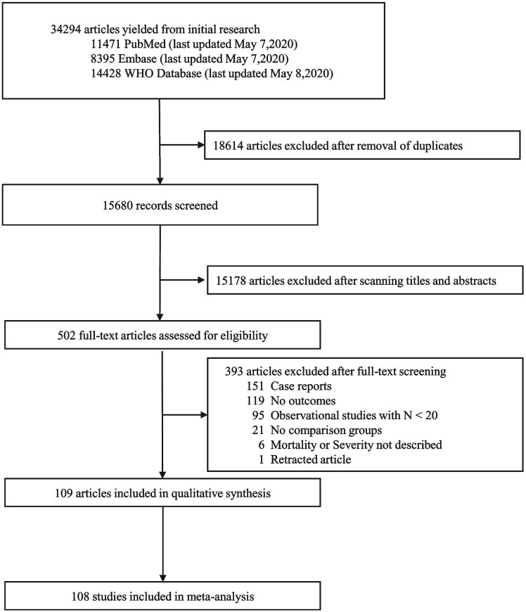
Fig 1
Full legend
Stages of the analysis process, assessing inclusion and exclusion criteria that amounted to the articles addressed in this review.
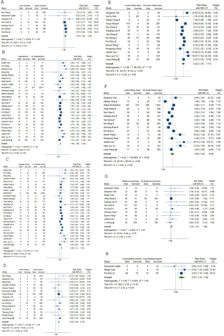
Fig 2
Full legend
Meta-analysis to assess risk of mortality for (A) smoking status. (B) hypertension. (C) diabetes mellitus. (D) COPD. (E) acute cardiac injury. (F) acute kidney injury. (G) bilateral lung involvement. (H) Lung consolidation. COPD = chronic obstructive pulmonary disease. !Shenzhen Third People’s Hospital, Shenzhen, China. #Tongji Hospital, Wuhan, China. $Wuhan Jinyintan Hospital, Wuhan, China. *Zhongnan Hospital of Wuhan University, Wuhan, China. %Tianyou Hospital, Wuhan, China. &Taizhou Public Health Medical Center, Zhejiang, China. +Chongqing Three Gorges Central Hospital, Chongqing, China. @Renmin Hospital of Wuhan University, Wuhan, China. ^General Hospital of Central Theater Command of People’s Liberation Army, Wuhan, China.
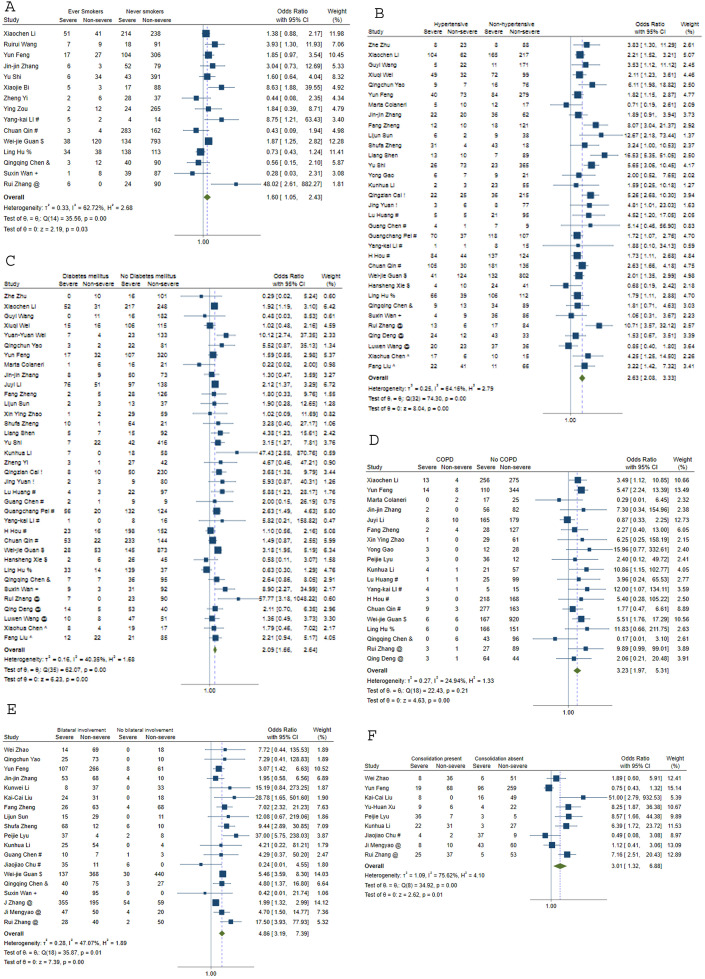
Fig 3
Full legend
Meta-analysis to assess odds of severe disease for (A) smoking status (B) hypertension (C) diabetes mellitus (D) COPD E. bilateral lung involvement (F) Lung consolidation. COPD = chronic obstructive pulmonary disease. !Shenzhen Third People’s Hospital, Shenzhen, China. # Tongji Hospital, Wuhan, China. $Wuhan Jinyintan Hospital, Wuhan, China. *Zhongnan Hospital of Wuhan University, Wuhan, China. %Tianyou Hospital, Wuhan, China. &Central hospital of Wuhan, Wuhan. +Chongqing Three Gorges Central Hospital, Chongqing, China. @Renmin Hospital of Wuhan University, Wuhan, China. ^General Hospital of Central Theater Command of People’s Liberation Army, Wuhan, China.
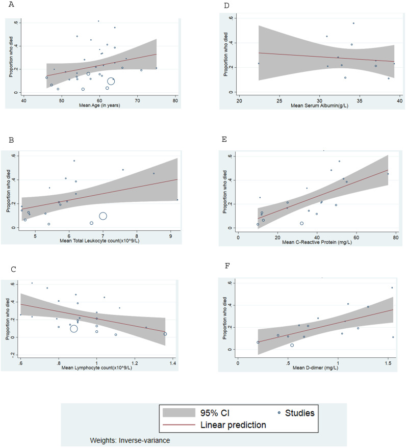
Fig 4
Full legend
Meta-regression plot showing the proportion increase in mortality among COVID-19 patients regressed against (A) mean age. (B) mean leukocyte count. (C) mean lymphocyte count. (D) mean serum albumin. (E) mean C-reactive protein. (F) mean D-dimer.
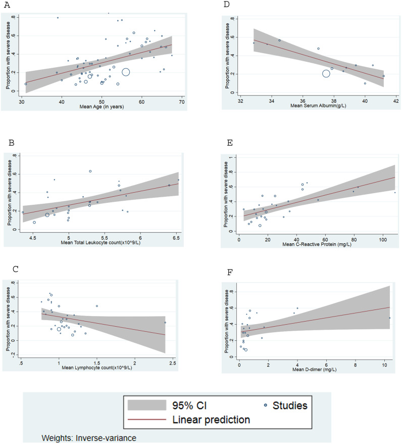
Fig 5
Full legend
Meta-regression plot shows the proportion increase in severity among COVID-19 patients regressed against (A) mean age. (B) mean leukocyte count. (C) mean lymphocyte count. (D) mean serum albumin. (E) mean C-reactive protein. (F) mean D-dimer.
References149
- 1.
WHO Director-General’s opening remarks at the media briefing on COVID-19–11 March 2020 [Internet]. [cited 2020 May 23]. https://www.who.int/dg/speeches/detail/who-director-general-s-opening-remarks-at-the-media-briefing-on-covid-19---11-march-2020
Not in our library
- 2.
WHO Timeline—COVID-19 [Internet]. [cited 2020 May 23]. https://www.who.int/news-room/detail/27-04-2020-who-timeline---covid-19
Not in our library
- 3.
Grasselli G, Pesenti A, Cecconi M. Critical Care Utilization for the COVID-19 Outbreak in Lombardy, Italy: Early Experience and Forecast During an Emergency Response. JAMA. 2020 4 28;323(16):1545–6.
Not in our library10.1001/jama.2020.4031 (opens in a new tab)
- 4.
Grasselli G, Zangrillo A, Zanella A, Antonelli M, Cabrini L, Castelli A, et al Baseline Characteristics and Outcomes of 1591 Patients Infected With SARS-CoV-2 Admitted to ICUs of the Lombardy Region, Italy. JAMA. 2020 4 28;323(16):1574–81.
Not in our library10.1001/jama.2020.5394 (opens in a new tab)
- 5.
Huang C, Wang Y, Li X, Ren L, Zhao J, Hu Y, et al Clinical features of patients infected with 2019 novel coronavirus in Wuhan, China. Lancet [Internet]. 2020; Available from: https://www.ncbi.nlm.nih.gov/pubmed/31986264
Not in our library10.1016/s0140-6736(20)30183-5 (opens in a new tab)
- 6.
Rhodes A, Ferdinande P, Flaatten H, Guidet B, Metnitz PG, Moreno RP. The variability of critical care bed numbers in Europe. Intensive Care Med. 2012 10;38(10):1647–53.
Not in our library10.1007/s00134-012-2627-8 (opens in a new tab)
- 7.
Maves RC, Downar J, Dichter JR, Hick JL, Devereaux A, Geiling JA, et al. Triage of Scarce Critical Care Resources in COVID-19 An Implementation Guide for Regional Allocation: An Expert Panel Report of the Task Force for Mass Critical Care and the American College of Chest Physicians. Chest. 2020 Apr 11;
Not in our library10.1016/j.chest.2020.03.063 (opens in a new tab)
- 8.
Pettilä V, Pettilä M, Sarna S, Voutilainen P, Takkunen O. Comparison of multiple organ dysfunction scores in the prediction of hospital mortality in the critically ill. Crit Care Med. 2002 8;30(8):1705–11.
Not in our library10.1097/00003246-200208000-00005 (opens in a new tab)
- 9.
- 10.
Glasziou PP, Sanders S, Hoffmann T. Waste in covid-19 research. BMJ [Internet]. 2020 5 12 [cited 2020 Jul 16];369 Available from: https://www.bmj.com/content/369/bmj.m1847
Not in our library10.1136/bmj.m1847 (opens in a new tab)
- 11.
Dekkers OM, Egger M, Altman DG, Vandenbroucke JP. Distinguishing Case Series From Cohort Studies. Annals of Internal Medicine. 2012 1 3;156(1_Part_1):37
Not in our library10.7326/0003-4819-156-1-201201030-00006 (opens in a new tab)
- 12.
Metlay JP, Waterer GW, Long AC, Anzueto A, Brozek J, Crothers K, et al Diagnosis and Treatment of Adults with Community-acquired Pneumonia. An Official Clinical Practice Guideline of the American Thoracic Society and Infectious Diseases Society of America. Am J Respir Crit Care Med. 2019 10 1;200(7):e45–67.
Not in our library10.1164/rccm.201908-1581st (opens in a new tab)
- 13.
Lin L, Li TS. Interpretation of “Guidelines for the Diagnosis and Treatment of Novel Coronavirus (2019-nCoV) Infection by the National Health Commission (Trial Version 5)”. Zhonghua Yi Xue Za Zhi. 2020;100(0):E001–E001.
Not in our library10.3760/cma.j.issn.0376-2491.2020.0001 (opens in a new tab)
- 14.
Covidence systematic review software, Veritas Health Innovation, Melbourne, Australia. Available at www.covidence.org.
Not in our library
- 15.
Qualtrics (2020) Qualtrics.com. Available at: http://www.qualtrics.com/ (accessed 24 march 2020).
Not in our library
- 16.
- 17.
Ottawa Hospital Research Institute [Internet]. [cited 2020 May 26]. http://www.ohri.ca/programs/clinical_epidemiology/oxford.asp
Not in our library
- 18.
StataCorp. 2019 Stata Statistical Software: Release 16. College Station, TX: StataCorp LLC.
Not in our library
- 19.
Yang X, Yu Y, Xu J, Shu H, Xia J, Liu H, et al Clinical course and outcomes of critically ill patients with SARS-CoV-2 pneumonia in Wuhan, China: a single-centered, retrospective, observational study. The Lancet Respiratory Medicine [Internet]. 2020; Available from: http://www.sciencedirect.com/science/article/pii/S2213260020300795
Not in our library10.1016/s2213-2600(20)30079-5 (opens in a new tab)
- 20.
Chen T, Wu D, Chen H, Yan W, Yang D, Chen G, et al Clinical characteristics of 113 deceased patients with coronavirus disease 2019: retrospective study. BMJ. 2020 3 26;m1091
Not in our library10.1136/bmj.m1091 (opens in a new tab)
- 21.
Tang N, Li D, Wang X, Sun Z. Abnormal coagulation parameters are associated with poor prognosis in patients with novel coronavirus pneumonia. Journal of Thrombosis and Haemostasis. 2020 4;18(4):844–7.
Not in our library10.1111/jth.14768 (opens in a new tab)
- 22.
Ruan Q, Yang K, Wang W, Jiang L, Song J. Clinical predictors of mortality due to COVID-19 based on an analysis of data of 150 patients from Wuhan, China. Intensive Care Med. 2020.
Not in our library10.1007/s00134-020-05991-x (opens in a new tab)
- 23.
Wu C, Chen X, Cai Y, Xia J, Zhou X, Xu S, et al Risk Factors Associated With Acute Respiratory Distress Syndrome and Death in Patients With Coronavirus Disease 2019 Pneumonia in Wuhan, China. JAMA Internal Medicine [Internet]. 2020 3 13 [cited 2020 May 25]; Available from: https://jamanetwork.com/journals/jamainternalmedicine/fullarticle/2763184
Not in our library10.1001/jamainternmed.2020.0994 (opens in a new tab)
- 24.
Li Y, Peng S, Li L, Wang Q, Ping W, Zhang N, et al Clinical and Transmission Characteristics of Covid-19—A Retrospective Study of 25 Cases from a Single Thoracic Surgery Department. Current Medical Science. 2020 4;40(2):295–300.
Not in our library10.1007/s11596-020-2176-2 (opens in a new tab)
- 25.
Deng Y, Liu W, Liu K, Fang Y-Y, Shang J, zhou L, et al Clinical characteristics of fatal and recovered cases of coronavirus disease 2019 (COVID-19) in Wuhan, China: a retrospective study. Chinese Medical Journal. 2020 3;1
Not in our library10.1097/cm9.0000000000000824 (opens in a new tab)
- 26.
- 27.
Zhou F, Yu T, Du R, Fan G, Liu Y, Liu Z, et al Clinical course and risk factors for mortality of adult inpatients with COVID-19 in Wuhan, China: a retrospective cohort study. The Lancet. 2020 3;395(10229):1054–62.
Not in our library10.1016/s0140-6736(20)30566-3 (opens in a new tab)
- 28.
Cao J, Tu W-J, Cheng W, Yu L, Liu Y-K, Hu X, et al Clinical Features and Short-term Outcomes of 102 Patients with Corona Virus Disease 2019 in Wuhan, China. Clinical Infectious Diseases [Internet]. 2020 4 2 [cited 2020 May 25]; Available from: https://academic.oup.com/cid/advance-article/doi/10.1093/cid/ciaa243/5814897
Not in our library10.1093/cid/ciaa243 (opens in a new tab)
- 29.
Du R-H, Liang L-R, Yang C-Q, Wang W, Cao T-Z, Li M, et al Predictors of mortality for patients with COVID-19 pneumonia caused by SARS-CoV-2: a prospective cohort study. European Respiratory Journal. 2020 5;55(5):2000524
Not in our library10.1183/13993003.00524-2020 (opens in a new tab)
- 30.
Tu W-J, Cao J, Yu L, Hu X, Liu Q. Clinicolaboratory study of 25 fatal cases of COVID-19 in Wuhan. Intensive Care Medicine [Internet]. 2020 4 6 [cited 2020 May 25]; Available from: http://link.springer.com/10.1007/s00134-020-06023-4
Not in our library10.1007/s00134-020-06023-4 (opens in a new tab)
- 31.
Wang L, He W, Yu X, Hu D, Bao M, Liu H, et al Coronavirus disease 2019 in elderly patients: Characteristics and prognostic factors based on 4-week follow-up. Journal of Infection. 2020 6;80(6):639–45.
Not in our library10.1016/j.jinf.2020.03.019 (opens in a new tab)
- 32.
Wang Y, Lu X, Chen H, Chen T, Su N, Huang F, et al Clinical Course and Outcomes of 344 Intensive Care Patients with COVID-19. American Journal of Respiratory and Critical Care Medicine [Internet]. 2020 4 8 [cited 2020 May 25]; Available from: https://www.atsjournals.org/doi/10.1164/rccm.202003-0736LE
Not in our library10.1164/rccm.202003-0736le (opens in a new tab)
- 33.
Dai M, Liu D, Liu M, Zhou F, Li G, Chen Z, et al Patients with cancer appear more vulnerable to SARS-COV-2: a multi-center study during the COVID-19 outbreak. Cancer Discovery. 2020 4 28;CD-20-0422.
Not in our library10.1158/2159-8290.cd-20-0422 (opens in a new tab)
- 34.
Ding M, Zhang Q, Li Q, Wu T, Huang Y. Correlation analysis of the severity and clinical prognosis of 32 cases of patients with COVID-19. Respiratory Medicine. 2020 6;167:105981
Not in our library10.1016/j.rmed.2020.105981 (opens in a new tab)
- 35.
Barrasa H, Rello J, Tejada S, Martín A, Balziskueta G, Vinuesa C, et al SARS-CoV-2 in Spanish Intensive Care Units: Early experience with 15-day survival in Vitoria. Anaesthesia Critical Care & Pain Medicine [Internet]. 2020 4 [cited 2020 May 25]; Available from: https://linkinghub.elsevier.com/retrieve/pii/S2352556820300643
Not in our library10.1016/j.accpm.2020.04.001 (opens in a new tab)
- 36.
Chen T, Dai Z, Mo P, Li X, Ma Z, Song S, et al Clinical Characteristics and Outcomes of Older Patients with Coronavirus Disease 2019 (COVID-19) in Wuhan, China: A Single-Centered, Retrospective Study. Newman A, editor. The Journals of Gerontology: Series A [Internet]. 2020 4 11 [cited 2020 May 25]; Available from: https://academic.oup.com/biomedgerontology/advance-article/doi/10.1093/gerona/glaa089/5819242
Not in our library10.1093/gerona/glaa089 (opens in a new tab)
- 37.
Trujillo H, Caravaca-Fontán F, Sevillano Á, Gutiérrez E, Caro J, Gutiérrez E, et al SARS-CoV-2 Infection in Hospitalized Patients With Kidney Disease. Kidney International Reports [Internet]. 2020 5 [cited 2020 May 25]; Available from: https://linkinghub.elsevier.com/retrieve/pii/S2468024920312274
Not in our library10.1016/j.ekir.2020.04.024 (opens in a new tab)
- 38.
Webb GJ, Moon AM, Barnes E, Barritt AS, Marjot T. Determining risk factors for mortality in liver transplant patients with COVID-19. The Lancet Gastroenterology & Hepatology [Internet]. 2020 4 [cited 2020 May 25]; Available from: https://linkinghub.elsevier.com/retrieve/pii/S2468125320301254
Not in our library10.1016/s2468-1253(20)30125-4 (opens in a new tab)
- 39.
Yao Q, Wang P, Wang X, Qie G, Meng M, Tong X, et al Retrospective study of risk factors for severe SARS-Cov-2 infections in hospitalized adult patients. Polish Archives of Internal Medicine [Internet]. 2020 4 24 [cited 2020 May 25]; Available from: https://www.mp.pl/paim/issue/article/15312
Not in our library10.20452/pamw.15312 (opens in a new tab)
- 40.
Wei F, Wang H, Huang H, Luo C, Zhou X, Xu N, et al Acute myocardial injury is common in patients with covid-19 and impairs their prognosis.: 6.
Not in our library10.1136/heartjnl-2020-317007 (opens in a new tab)
- 41.
Tomlins J, Hamilton F, Gunning S, Sheehy C, Moran E, MacGowan A. Clinical features of 95 sequential hospitalised patients with novel coronavirus 2019 disease (COVID-19), the first UK cohort. Journal of Infection [Internet]. 2020 4 [cited 2020 May 25]; Available from: https://linkinghub.elsevier.com/retrieve/pii/S0163445320302322
Not in our library10.1016/j.jinf.2020.04.020 (opens in a new tab)
- 42.
Yang X, Yang Q, Wang Y, Wu Y, Xu J, Yu Y, et al Thrombocytopenia and its association with mortality in patients with COVID-19. Journal of Thrombosis and Haemostasis [Internet]. 2020 5 4 [cited 2020 May 25]; Available from: https://onlinelibrary.wiley.com/doi/abs/10.1111/jth.14848
Not in our library10.1111/jth.14848 (opens in a new tab)
- 43.
Yang R, Gui X, Zhang Y, Xiong Y. The role of essential organ-based comorbidities in the prognosis of COVID-19 infection patients. Expert Review of Respiratory Medicine. 2020 4 28;1–4.
Not in our library10.1080/17476348.2020.1761791 (opens in a new tab)
- 44.
Richardson S, Hirsch JS, Narasimhan M, Crawford JM, McGinn T, Davidson KW, et al Presenting Characteristics, Comorbidities, and Outcomes Among 5700 Patients Hospitalized With COVID-19 in the New York City Area. JAMA [Internet]. 2020 4 22 [cited 2020 May 25]; Available from: https://jamanetwork.com/journals/jama/fullarticle/2765184
Not in our library10.1001/jama.2020.6775 (opens in a new tab)
- 45.
- 46.
- 47.
Singh S, Khan A. Clinical Characteristics and Outcomes of COVID-19 Among Patients with Pre-Existing Liver Disease in United States: A Multi-Center Research Network Study. Gastroenterology [Internet]. 2020 5 [cited 2020 May 25]; Available from: https://linkinghub.elsevier.com/retrieve/pii/S0016508520305850
Not in our library
- 48.
Zangrillo A, Beretta L, Scandroglio AM, Monti G, Fominskiy E, Colombo S, et al Characteristics, treatment, outcomes and cause of death of invasively ventilated patients with COVID-19 ARDS in Milan, Italy. Critical Care and Resuscitation.: 12.
Not in our library10.1016/s1441-2772(23)00387-3 (opens in a new tab)
- 49.
Yan Y, Yang Y, Wang F, Ren H, Zhang S, Shi X, et al Clinical characteristics and outcomes of patients with severe covid-19 with diabetes. BMJ Open Diabetes Research & Care. 2020 4;8(1):e001343.
Not in our library10.1136/bmjdrc-2020-001343 (opens in a new tab)
- 50.
Wang K, Zuo P, Liu Y, Zhang M, Zhao X, Xie S, et al Clinical and laboratory predictors of in-hospital mortality in patients with COVID-19: a cohort study in Wuhan, China. Clin Infect Dis. 2020 5 3;
Not in our library10.1093/cid/ciaa538 (opens in a new tab)
- 51.
Wang K, Zhang Z, Yu M, Tao Y, Xie M. 15-day mortality and associated risk factors for hospitalized patients with COVID-19 in Wuhan, China: an ambispective observational cohort study. Intensive Care Medicine [Internet]. 2020 4 23 [cited 2020 May 25]; Available from: http://link.springer.com/10.1007/s00134-020-06047-w
Not in our library10.1007/s00134-020-06047-w (opens in a new tab)
- 52.
Zhang L, Yan X, Fan Q, Liu H, Liu X, Liu Z, et al D-dimer levels on admission to predict in-hospital mortality in patients with Covid-19. Journal of Thrombosis and Haemostasis. 2020;18(6):1324–9.
Not in our library10.1111/jth.14859 (opens in a new tab)
- 53.
- 54.
Liu Y, Sun W, Guo Y, Chen L, Zhang L, Zhao S, et al Association between platelet parameters and mortality in coronavirus disease 2019: Retrospective cohort study. Platelets. 2020 5 18;31(4):490–6.
Not in our library10.1080/09537104.2020.1754383 (opens in a new tab)
- 55.
Liu Y, Du X, Chen J, Jin Y, Peng L, Wang HHX, et al Neutrophil-to-lymphocyte ratio as an independent risk factor for mortality in hospitalized patients with COVID-19. Journal of Infection [Internet]. 2020 4 [cited 2020 May 25]; Available from: https://linkinghub.elsevier.com/retrieve/pii/S0163445320302085
Not in our library10.1016/j.jinf.2020.04.002 (opens in a new tab)
- 56.
Li J, Wang X, Chen J, Zhang H, Deng A. Association of Renin-Angiotensin System Inhibitors With Severity or Risk of Death in Patients With Hypertension Hospitalized for Coronavirus Disease 2019 (COVID-19) Infection in Wuhan, China. JAMA Cardiology [Internet]. 2020 4 23 [cited 2020 May 25]; Available from: https://jamanetwork.com/journals/jamacardiology/fullarticle/2765049
Not in our library10.1001/jamacardio.2020.1624 (opens in a new tab)
- 57.
Nikpouraghdam M, Jalali Farahani A, Alishiri G, Heydari S, Ebrahimnia M, Samadinia H, et al Epidemiological characteristics of coronavirus disease 2019 (COVID-19) patients in IRAN: A single center study. Journal of Clinical Virology. 2020 6;127:104378
Not in our library10.1016/j.jcv.2020.104378 (opens in a new tab)
- 58.
Zhang J, Wang X, Jia X, Li J, Hu K, Chen G, et al Risk factors for disease severity, unimprovement, and mortality in COVID-19 patients in Wuhan, China. Clinical Microbiology and Infection. 2020 6;26(6):767–72.
Not in our library10.1016/j.cmi.2020.04.012 (opens in a new tab)
- 59.
Mehta V, Goel S, Kabarriti R, Cole D, Goldfinger M, Acuna-Villaorduna A, et al Case Fatality Rate of Cancer Patients with COVID-19 in a New York Hospital System. Cancer Discovery. 2020 5 1;CD-20-0516.
Not in our library10.1158/2159-8290.cd-20-0516 (opens in a new tab)
- 60.
Zou X, Li S, Fang M, Hu M, Bian Y, Ling J, et al Acute Physiology and Chronic Health Evaluation II Score as a Predictor of Hospital Mortality in Patients of Coronavirus Disease 2019: Critical Care Medicine. 2020 5;1.
Not in our library10.1097/ccm.0000000000004411 (opens in a new tab)
- 61.
Guan W, Ni Z, Hu Y, Liang W, Ou C, He J, et al Clinical Characteristics of Coronavirus Disease 2019 in China. New England Journal of Medicine [Internet]. 2020; Available from:
Not in our library10.1056/nejmoa2002032 (opens in a new tab)
- 62.
Xu Y-H, Dong J-H, An W, Lv X-Y, Yin X-P, Zhang J-Z, et al Clinical and computed tomographic imaging features of Novel Coronavirus Pneumonia caused by SARS-CoV-2. Journal of Infection [Internet]. 2020; Available from: http://www.sciencedirect.com/science/article/pii/S0163445320301006
Not in our library10.1016/j.jinf.2020.02.017 (opens in a new tab)
- 63.
Tian S, Hu N, Lou J, Chen K, Kang X, Xiang Z, et al Characteristics of COVID-19 infection in Beijing. Journal of Infection. 2020 4;80(4):401–6.
Not in our library10.1016/j.jinf.2020.02.018 (opens in a new tab)
- 64.
Li K, Wu J, Wu F, Guo D, Chen L, Fang Z, et al The Clinical and Chest CT Features Associated With Severe and Critical COVID-19 Pneumonia. Investigative Radiology. 2020 6;55(6):327–31.
Not in our library10.1097/rli.0000000000000672 (opens in a new tab)
- 65.
Zhao W, Zhong Z, Xie X, Yu Q, Liu J. Relation Between Chest CT Findings and Clinical Conditions of Coronavirus Disease (COVID-19) Pneumonia: A Multicenter Study. AJR Am J Roentgenol. 2020;1–6.
Not in our library10.2214/ajr.20.22976 (opens in a new tab)
- 66.
Han H, Yang L, Liu R, Liu F, Wu K, Li J, et al Prominent changes in blood coagulation of patients with SARS-CoV-2 infection. Clinical Chemistry and Laboratory Medicine (CCLM) [Internet]. 2020 3 16 [cited 2020 May 25];0(0). Available from: https://www.degruyter.com/view/journals/cclm/ahead-of-print/article-10.1515-cclm-2020-0188/article-10.1515-cclm-2020-0188.xml
Not in our library10.1515/cclm-2020-0188 (opens in a new tab)
- 67.
Qian G-Q, Yang N-B, Ding F, Ma AHY, Wang Z-Y, Shen Y-F, et al Epidemiologic and Clinical Characteristics of 91 Hospitalized Patients with COVID-19 in Zhejiang, China: A retrospective, multi-centre case series. QJM. 2020 3 17;
Not in our library10.1093/qjmed/hcaa089 (opens in a new tab)
- 68.
Qu R, Ling Y, Zhang Y, Wei L, Chen X, Li X, et al Platelet-to-lymphocyte ratio is associated with prognosis in patients with Corona Virus Disease-19. Journal of Medical Virology [Internet]. 2020 3 17 [cited 2020 May 25]; Available from: https://onlinelibrary.wiley.com/doi/abs/10.1002/jmv.25767
Not in our library10.1002/jmv.25767 (opens in a new tab)
- 69.
Gao Y, Li T, Han M, Li X, Wu D, Xu Y, et al Diagnostic Utility of Clinical Laboratory Data Determinations for Patients with the Severe COVID-19. Journal of Medical Virology [Internet]. 2020 3 17 [cited 2020 May 25]; Available from: https://onlinelibrary.wiley.com/doi/abs/10.1002/jmv.25770
Not in our library10.1002/jmv.25770 (opens in a new tab)
- 70.
Liu K-C, Xu P, Lv W-F, Qiu X-H, Yao J-L, Gu J-F, et al CT manifestations of coronavirus disease-2019: A retrospective analysis of 73 cases by disease severity. European Journal of Radiology. 2020 5;126:108941
Not in our library10.1016/j.ejrad.2020.108941 (opens in a new tab)
- 71.
Wan S, Xiang Y, Fang W, Zheng Y, Li B, Hu Y, et al Clinical features and treatment of COVID-19 patients in northeast Chongqing. Journal of Medical Virology [Internet]. 2020 4 1 [cited 2020 May 25]; Available from: http://doi.wiley.com/10.1002/jmv.25783
Not in our library10.1002/jmv.25783 (opens in a new tab)
- 72.
- 73.
Li K, Fang Y, Li W, Pan C, Qin P, Zhong Y, et al CT image visual quantitative evaluation and clinical classification of coronavirus disease (COVID-19). European Radiology [Internet]. 2020 3 25 [cited 2020 May 25]; Available from: http://link.springer.com/10.1007/s00330-020-06817-6
Not in our library10.1007/s00330-020-06817-6 (opens in a new tab)
- 74.
Qin C, Zhou L, Hu Z, Zhang S, Yang S, Tao Y, et al Dysregulation of Immune Response in Patients With Coronavirus 2019 (COVID-19) in Wuhan, China. Clin Infect Dis [Internet]. [cited 2020 Jun 6]; Available from: https://academic.oup.com/cid/advance-article/doi/10.1093/cid/ciaa248/5803306
Not in our library10.1093/cid/ciaa248 (opens in a new tab)
- 75.
Huang L, Han R, Ai T, Yu P, Kang H, Tao Q, et al Serial Quantitative Chest CT Assessment of COVID-19: Deep-Learning Approach. Radiology: Cardiothoracic Imaging. 2020 4 1;2(2):e200075.
Not in our library10.1148/ryct.2020200075 (opens in a new tab)
- 76.
Zhang J-J, Dong X, Cao Y-Y, Yuan Y-D, Yang Y-B, Yan Y-Q, et al Clinical characteristics of 140 patients infected by SARS-CoV-2 in Wuhan, China. Allergy. 2020;
Not in our library10.1111/all.14238 (opens in a new tab)
- 77.
Chen G, Wu D, Guo W, Cao Y, Huang D, Wang H, et al Clinical and immunological features of severe and moderate coronavirus disease 2019. Journal of Clinical Investigation. 2020 4 13;130(5):2620–9.
Not in our library10.1172/jci137244 (opens in a new tab)
- 78.
Chu J, Yang N, Wei Y, Yue H, Zhang F, Zhao J, et al Clinical characteristics of 54 medical staff with COVID-19: A retrospective study in a single center in Wuhan, China. Journal of Medical Virology [Internet]. 2020 4 6 [cited 2020 May 25]; Available from: https://onlinelibrary.wiley.com/doi/abs/10.1002/jmv.25793
Not in our library10.1002/jmv.25793 (opens in a new tab)
- 79.
Yuan J, Zou R, Zeng L, Kou S, Lan J, Li X, et al The correlation between viral clearance and biochemical outcomes of 94 COVID-19 infected discharged patients. Inflammation Research. 2020 6;69(6):599–606.
Not in our library10.1007/s00011-020-01342-0 (opens in a new tab)
- 80.
Wang L, Li X, Chen H, Yan S, Li D, Li Y, et al Coronavirus Disease 19 Infection Does Not Result in Acute Kidney Injury: An Analysis of 116 Hospitalized Patients from Wuhan, China. American Journal of Nephrology. 2020;51(5):343–8.
Not in our library10.1159/000507471 (opens in a new tab)
- 81.
Han H, Xie L, Liu R, Yang J, Liu F, Wu K, et al Analysis of heart injury laboratory parameters in 273 COVID-19 patients in one hospital in Wuhan, China. Journal of Medical Virology [Internet]. 2020 4 15 [cited 2020 May 25]; Available from: http://doi.wiley.com/10.1002/jmv.25809
Not in our library10.1002/jmv.25809 (opens in a new tab)
- 82.
Xie H, Zhao J, Lian N, Lin S, Xie Q, Zhuo H. Clinical characteristics of non-ICU hospitalized patients with coronavirus disease 2019 and liver injury: A retrospective study. Liver International [Internet]. 2020 4 12 [cited 2020 May 25]; Available from: http://doi.wiley.com/10.1111/liv.14449
Not in our library10.1111/liv.14449 (opens in a new tab)
- 83.
Cai Q, Huang D, Ou P, Yu H, Zhu Z, Xia Z, et al COVID-19 in a designated infectious diseases hospital outside Hubei Province, China. Allergy [Internet]. 2020 4 17 [cited 2020 May 25]; Available from: https://onlinelibrary.wiley.com/doi/abs/10.1111/all.14309
Not in our library10.1111/all.14309 (opens in a new tab)
- 84.
Zhang Y, Zheng L, Liu L, Zhao M, Xiao J, Zhao Q. Liver impairment in COVID-19 patients: A retrospective analysis of 115 cases from a single centre in Wuhan city, China. Liver International [Internet]. 2020 4 28 [cited 2020 May 25]; Available from: https://onlinelibrary.wiley.com/doi/abs/10.1111/liv.14455
Not in our library10.1111/liv.14455 (opens in a new tab)
- 85.
Tan C, Huang Y, Shi F, Tan K, Ma Q, Chen Y, et al C-reactive protein correlates with computed tomographic findings and predicts severe COVID-19 early. J Med Virol [Internet]. 2020 4 25 [cited 2020 Jun 5]; Available from: https://www.ncbi.nlm.nih.gov/pmc/articles/PMC7262341/
Not in our library10.1002/jmv.25871 (opens in a new tab)
- 86.
Chen X, Yang Y, Huang M, Liu L, Zhang X, Xu J, et al Differences between COVID-19 and suspected then confirmed SARS-CoV-2-negative pneumonia: a retrospective study from a single center. Journal of Medical Virology [Internet]. 2020 4 1 [cited 2020 May 25]; Available from: http://doi.wiley.com/10.1002/jmv.25810
Not in our library10.1002/jmv.25810 (opens in a new tab)
- 87.
Zheng F, Tang W, Li H. Clinical characteristics of 161 cases of corona virus disease 2019 (COVID-19) in Changsha.: 7.
Not in our library10.26355/eurrev_202003_20711 (opens in a new tab)
- 88.
Deng Q, Hu B, Zhang Y, Wang H, Zhou X, Hu W, et al Suspected myocardial injury in patients with COVID-19: Evidence from front-line clinical observation in Wuhan, China. International Journal of Cardiology [Internet]. 2020 4 [cited 2020 May 25]; Available from: https://linkinghub.elsevier.com/retrieve/pii/S0167527320311153
Not in our library10.1016/j.ijcard.2020.03.087 (opens in a new tab)
- 89.
Li H, Xiang X, Ren H, Xu L, Zhao L, Chen X, et al Serum Amyloid A is a biomarker of severe Coronavirus Disease and poor prognosis. Journal of Infection. 2020 6;80(6):646–55.
Not in our library10.1016/j.jinf.2020.03.035 (opens in a new tab)
- 90.
Zou Y, Guo H, Zhang Y, Zhang Z, Liu Y, Wang J, et al Analysis of coagulation parameters in patients with COVID-19 in Shanghai, China. BioScience Trends [Internet]. 2020 [cited 2020 May 25]; Available from: https://www.jstage.jst.go.jp/article/bst/advpub/0/advpub_2020.03086/_article
Not in our library10.5582/bst.2020.03086 (opens in a new tab)
- 91.
Shi F, Wu T, Zhu X, Ge Y, Zeng X, Chi Y, et al Association of viral load with serum biomakers among COVID-19 cases. Virology. 2020 7;546:122–6.
Not in our library10.1016/j.virol.2020.04.011 (opens in a new tab)
- 92.
Wang G, Wu C, Zhang Q, Wu F, Yu B, Lv J, et al C-Reactive Protein Level May Predict the Risk of COVID-19 Aggravation. Open Forum Infectious Diseases [Internet]. 2020 5 1 [cited 2020 May 25];7(5). Available from: https://academic.oup.com/ofid/article/doi/10.1093/ofid/ofaa153/5826961
Not in our library10.1093/ofid/ofaa153 (opens in a new tab)
- 93.
Ji M, Yuan L, Shen W, Lv J, Li Y, Li M, et al Characteristics of disease progress in patients with coronavirus disease 2019 in Wuhan, China. Epidemiology and Infection [Internet]. 2020 [cited 2020 May 25];148 Available from: https://www.cambridge.org/core/product/identifier/S0950268820000977/type/journal_article
Not in our library10.1017/s0950268820000977 (opens in a new tab)
- 94.
Shen L, Li S, Zhu Y, Zhao J, Tang X, Li H, et al Clinical and laboratory-derived parameters of 119 hospitalized patients with coronavirus disease 2019 in Xiangyang, Hubei Province, China. Journal of Infection [Internet]. 2020 4 [cited 2020 May 25]; Available from: https://linkinghub.elsevier.com/retrieve/pii/S0163445320301663
Not in our library10.1016/j.jinf.2020.03.038 (opens in a new tab)
- 95.
Yang F, Shi S, Zhu J, Shi J, Dai K, Chen X. Clinical characteristics and outcomes of cancer patients with COVID-19. Journal of Medical Virology [Internet]. 2020 5 5 [cited 2020 May 25]; Available from: https://onlinelibrary.wiley.com/doi/abs/10.1002/jmv.25972
Not in our library10.1002/jmv.25972 (opens in a new tab)
- 96.
Ma J, Yin J, Qian Y, Wu Y. Clinical characteristics and prognosis in cancer patients with COVID-19: A single center’s retrospective study. Journal of Infection [Internet]. 2020 4 [cited 2020 May 25]; Available from: https://linkinghub.elsevier.com/retrieve/pii/S0163445320302140
Not in our library10.1016/j.jinf.2020.04.006 (opens in a new tab)
- 97.
- 98.
Colaneri M, Sacchi P, Zuccaro V, Biscarini S, Sachs M, Roda S, et al Clinical characteristics of coronavirus disease (COVID-19) early findings from a teaching hospital in Pavia, North Italy, 21 to 28 February 2020. Eurosurveillance [Internet]. 2020 4 23 [cited 2020 May 25];25(16). Available from: https://www.eurosurveillance.org/content/10.2807/1560-7917.ES.2020.25.16.2000460
Not in our library10.2807/1560-7917.es.2020.25.16.2000460 (opens in a new tab)
- 99.
- 100.
Chen L, Li Q, Zheng D, Jiang H, Wei Y, Zou L, et al Clinical Characteristics of Pregnant Women with Covid-19 in Wuhan, China. New England Journal of Medicine [Internet]. 2020 4 17 [cited 2020 May 25]; Available from: http://www.nejm.org/doi/10.1056/NEJMc2009226
Not in our library10.1056/nejmc2009226 (opens in a new tab)
- 101.
Sun L, Shen L, Fan J, Gu F, Hu M, An Y, et al Clinical Features of Patients with Coronavirus Disease 2019 (COVID-19) from a Designated Hospital in Beijing, China. Journal of Medical Virology [Internet]. 2020 5 5 [cited 2020 May 25]; Available from: https://onlinelibrary.wiley.com/doi/abs/10.1002/jmv.25966
Not in our library10.1002/jmv.25966 (opens in a new tab)
- 102.
Zhu Z, Cai T, Fan L, Lou K, Hua X, Huang Z, et al Clinical value of immune-inflammatory parameters to assess the severity of coronavirus disease 2019. International Journal of Infectious Diseases. 2020 6;95:332–9.
Not in our library10.1016/j.ijid.2020.04.041 (opens in a new tab)
- 103.
Liu Y, Liao W, Wan L, Xiang T, Zhang W. Correlation Between Relative Nasopharyngeal Virus RNA Load and Lymphocyte Count Disease Severity in Patients with COVID-19. Viral Immunology [Internet]. 2020 4 10 [cited 2020 May 25]; Available from: https://www.liebertpub.com/doi/10.1089/vim.2020.0062
Not in our library10.1089/vim.2020.0062 (opens in a new tab)
- 104.
Feng Y, Ling Y, Bai T, Xie Y, Huang J, Li J, et al COVID-19 with Different Severity: A Multi-center Study of Clinical Features. American Journal of Respiratory and Critical Care Medicine [Internet]. 2020 4 10 [cited 2020 May 25]; Available from: https://www.atsjournals.org/doi/10.1164/rccm.202002-0445OC
Not in our library10.1164/rccm.202002-0445oc (opens in a new tab)
- 105.
Zhang R, Ouyang H, Fu L, Wang S, Han J, Huang K, et al CT features of SARS-CoV-2 pneumonia according to clinical presentation: a retrospective analysis of 120 consecutive patients from Wuhan city. European Radiology [Internet]. 2020 4 11 [cited 2020 May 25]; Available from: http://link.springer.com/10.1007/s00330-020-06854-1
Not in our library10.1007/s00330-020-06854-1 (opens in a new tab)
- 106.
Chen X, Zhao B, Qu Y, Chen Y, Xiong J, Feng Y, et al Detectable serum SARS-CoV-2 viral load (RNAaemia) is closely correlated with drastically elevated interleukin 6 (IL-6) level in critically ill COVID-19 patients. Clinical Infectious Diseases [Internet]. 2020 4 17 [cited 2020 May 25]; Available from: https://academic.oup.com/cid/advance-article/doi/10.1093/cid/ciaa449/5821311
Not in our library10.1093/cid/ciaa449 (opens in a new tab)
- 107.
Wei X, Su J, Yang K, Wei J, Wan H, Cao X, et al Elevations of serum cancer biomarkers correlate with severity of COVID-19. Journal of Medical Virology [Internet]. 2020 4 29 [cited 2020 May 25]; Available from: https://onlinelibrary.wiley.com/doi/abs/10.1002/jmv.25957
Not in our library10.1002/jmv.25957 (opens in a new tab)
- 108.
Zheng Y, Xiong C, Liu Y, Qian X, Tang Y, Liu L, et al Epidemiological and clinical characteristics analysis of COVID-19 in the surrounding areas of Wuhan, Hubei Province in 2020. Pharmacol Res. 2020 7;157:104821
Not in our library10.1016/j.phrs.2020.104821 (opens in a new tab)
- 109.
- 110.
Yao Z, Zheng Z, Wu K, Junhua Z. Immune environment modulation in pneumonia patients caused by coronavirus: SARS-CoV, MERS-CoV and SARS-CoV-2. Aging. 2020 5 2;12(9):7639–51.
Not in our library10.18632/aging.103101 (opens in a new tab)
- 111.
Lei F, Liu Y, Zhou F, Qin J, Zhang P, Zhu L, et al Longitudinal association between markers of liver injury and mortality in COVID-19 in China. Hepatology [Internet]. 2020 5 2 [cited 2020 May 25]; Available from: https://onlinelibrary.wiley.com/doi/abs/10.1002/hep.31301
Not in our library10.1002/hep.31301 (opens in a new tab)
- 112.
Lu L, Xiong W, Liu D, Liu J, Yang D, Li N, et al New onset acute symptomatic seizure and risk factors in coronavirus disease 2019: A retrospective multicenter study. Epilepsia [Internet]. 2020 5 2 [cited 2020 May 25]; Available from: https://onlinelibrary.wiley.com/doi/abs/10.1111/epi.16524
Not in our library10.1111/epi.16524 (opens in a new tab)
- 113.
Yang Y, Shen C, Li J, Yuan J, Wei J, Huang F, et al Plasma IP-10 and MCP-3 levels are highly associated with disease severity and predict the progression of COVID-19. Journal of Allergy and Clinical Immunology [Internet]. 2020 4 [cited 2020 May 25]; Available from: https://linkinghub.elsevier.com/retrieve/pii/S0091674920305765
Not in our library10.1016/j.jaci.2020.04.027 (opens in a new tab)
- 114.
Bi X, Su Z, Yan H, Du J, Wang J, Chen L, et al Prediction of severe illness due to COVID-19 based on an analysis of initial Fibrinogen to Albumin Ratio and Platelet count. Platelets. 2020 5 5;1–6.
Not in our library10.1080/09537104.2020.1760230 (opens in a new tab)
- 115.
Liu F, Li L, Xu M, Wu J, Luo D, Zhu Y, et al Prognostic value of interleukin-6, C-reactive protein, and procalcitonin in patients with COVID-19. Journal of Clinical Virology. 2020 6;127:104370
Not in our library10.1016/j.jcv.2020.104370 (opens in a new tab)
- 116.
Pei G, Zhang Z, Peng J, Liu L, Zhang C, Yu C, et al Renal Involvement and Early Prognosis in Patients with COVID-19 Pneumonia. Journal of the American Society of Nephrology. 2020 4 28;ASN.2020030276.
Not in our library10.1681/asn.2020030276 (opens in a new tab)
- 117.
Hu L, Chen S, Fu Y, Gao Z, Long H, Wang J, et al Risk Factors Associated with Clinical Outcomes in 323 COVID-19 Hospitalized Patients in Wuhan, China. Clinical Infectious Diseases [Internet]. 2020 5 3 [cited 2020 May 25]; Available from: https://academic.oup.com/cid/advance-article/doi/10.1093/cid/ciaa539/5828282
Not in our library10.1093/cid/ciaa539 (opens in a new tab)
- 118.
- 119.
Wei Y-Y, Wang R-R, Zhang D-W, Tu Y-H, Chen C-S, Ji S, et al Risk factors for severe COVID-19: Evidence from 167 hospitalized patients in Anhui, China. Journal of Infection [Internet]. 2020 4 [cited 2020 May 25]; Available from: https://linkinghub.elsevier.com/retrieve/pii/S016344532030219X
Not in our library10.1016/j.jinf.2020.04.010 (opens in a new tab)
- 120.
Li X, Xu S, Yu M, Wang K, Tao Y, Zhou Y, et al Risk factors for severity and mortality in adult COVID-19 inpatients in Wuhan. Journal of Allergy and Clinical Immunology [Internet]. 2020 4 [cited 2020 May 25]; Available from: https://linkinghub.elsevier.com/retrieve/pii/S0091674920304954
Not in our library10.1016/j.jaci.2020.04.006 (opens in a new tab)
- 121.
Zheng Y, Zhang Y, Chi H, Chen S, Peng M, Luo L, et al The hemocyte counts as a potential biomarker for predicting disease progression in COVID-19: a retrospective study. Clinical Chemistry and Laboratory Medicine (CCLM) [Internet]. 2020 4 29 [cited 2020 May 25];0(0). Available from: https://www.degruyter.com/view/journals/cclm/ahead-of-print/article-10.1515-cclm-2020-0377/article-10.1515-cclm-2020-0377.xml
Not in our library10.1515/cclm-2020-0377 (opens in a new tab)
- 122.
Wang F, Hou H, Luo Y, Tang G, Wu S, Huang M, et al The laboratory tests and host immunity of COVID-19 patients with different severity of illness. JCI Insight [Internet]. 2020 5 21 [cited 2020 May 25];5(10). Available from: https://insight.jci.org/articles/view/137799
Not in our library10.1172/jci.insight.137799 (opens in a new tab)
- 123.
Lyu P, Liu X, Zhang R, Shi L, Gao J. The performance of chest CT in evaluating the clinical severity of COVID-19 pneumonia: identifying critical cases based on CT characteristics. Investigative Radiology. 2020 4;1
Not in our library10.1097/rli.0000000000000689 (opens in a new tab)
- 124.
Liu R, Ma Q, Han H, Su H, Liu F, Wu K, et al The value of urine biochemical parameters in the prediction of the severity of coronavirus disease 2019. Clinical Chemistry and Laboratory Medicine (CCLM) [Internet]. 2020 4 14 [cited 2020 May 25];0(0). Available from: https://www.degruyter.com/view/journals/cclm/ahead-of-print/article-10.1515-cclm-2020-0220/article-10.1515-cclm-2020-0220.xml
Not in our library10.1515/cclm-2020-0220 (opens in a new tab)
- 125.
Hou H, Zhang B, Huang H, Luo Y, Wu S, Tang G, et al Using IL-2R/lymphocytes for predicting the clinical progression of patients with COVID-19. Clinical & Experimental Immunology [Internet]. 2020 5 15 [cited 2020 May 25]; Available from: https://onlinelibrary.wiley.com/doi/abs/10.1111/cei.13450
Not in our library10.1111/cei.13450 (opens in a new tab)
- 126.
Zheng S, Fan J, Yu F, Feng B, Lou B, Zou Q, et al Viral load dynamics and disease severity in patients infected with SARS-CoV-2 in Zhejiang province, China, January-March 2020: retrospective cohort study. BMJ. 2020 4 21;m1443
Not in our library10.1136/bmj.m1443 (opens in a new tab)
- 127.
Huang I, Lim MA, Pranata R. Diabetes mellitus is associated with increased mortality and severity of disease in COVID-19 pneumonia—A systematic review, meta-analysis, and meta-regression. Diabetes & Metabolic Syndrome: Clinical Research & Reviews. 2020 7 1;14(4):395–403.
Not in our library10.1016/j.dsx.2020.04.018 (opens in a new tab)
- 128.
Pranata R, Huang I, Lim MA, Wahjoepramono EJ, July J. Impact of cerebrovascular and cardiovascular diseases on mortality and severity of COVID-19–systematic review, meta-analysis, and meta-regression. Journal of Stroke and Cerebrovascular Diseases. 2020 8 1;29(8):104949.
Not in our library10.1016/j.jstrokecerebrovasdis.2020.104949 (opens in a new tab)
- 129.
Yeh J-J, Lin C-L, Kao C-H. Relationship between pneumonia and cardiovascular diseases: A retrospective cohort study of the general population. Eur J Intern Med. 2019;59:39–45.
Not in our library10.1016/j.ejim.2018.08.003 (opens in a new tab)
- 130.
Wang D, Hu B, Hu C, Zhu F, Liu X, Zhang J, et al Clinical Characteristics of 138 Hospitalized Patients With 2019 Novel Coronavirus-Infected Pneumonia in Wuhan, China. JAMA. 2020;
Not in our library10.1001/jama.2020.1585 (opens in a new tab)
- 131.
Yu C-M. Cardiovascular complications of severe acute respiratory syndrome. Postgraduate Medical Journal. 2006 2 1;82(964):140–4.
Not in our library10.1136/pgmj.2005.037515 (opens in a new tab)
- 132.
Oudit GY, Kassiri Z, Jiang C, Liu PP, Poutanen SM, Penninger JM, et al SARS-coronavirus modulation of myocardial ACE2 expression and inflammation in patients with SARS. Eur J Clin Invest. 2009 7;39(7):618–25.
Not in our library10.1111/j.1365-2362.2009.02153.x (opens in a new tab)
- 133.
- 134.
Singh S, Sharma A, Arora SK. High Producer Haplotype (CAG) of -863C/A, -308G/A and -238G/A Polymorphisms in the Promoter Region of TNF-α Gene Associate with Enhanced Apoptosis of Lymphocytes in HIV-1 Subtype C Infected Individuals from North India. PLoS One [Internet]. 2014 5 16 [cited 2020 Jun 4];9(5). Available from: https://www.ncbi.nlm.nih.gov/pmc/articles/PMC4024031/
Not in our library10.1371/journal.pone.0098020 (opens in a new tab)
- 135.
Liao Y-C, Liang W-G, Chen F-W, Hsu J-H, Yang J-J, Chang M-S. IL-19 induces production of IL-6 and TNF-alpha and results in cell apoptosis through TNF-alpha. J Immunol. 2002 10 15;169(8):4288–97.
Not in our library10.4049/jimmunol.169.8.4288 (opens in a new tab)
- 136.
Aggarwal S, Gollapudi S, Gupta S. Increased TNF-alpha-induced apoptosis in lymphocytes from aged humans: changes in TNF-alpha receptor expression and activation of caspases. J Immunol. 1999 2 15;162(4):2154–61.
Not in our library
- 137.
Simon L, Gauvin F, Amre DK, Saint-Louis P, Lacroix J. Serum procalcitonin and C-reactive protein levels as markers of bacterial infection: a systematic review and meta-analysis. Clin Infect Dis. 2004 7 15;39(2):206–17.
Not in our library10.1086/421997 (opens in a new tab)
- 138.
Levi M, van der Poll T. Coagulation and sepsis. Thrombosis Research. 2017 1;149:38–44.
Not in our library10.1016/j.thromres.2016.11.007 (opens in a new tab)
- 139.
- 140.
Gupta N, Zhao Y-Y, Evans CE. The stimulation of thrombosis by hypoxia. Thrombosis Research. 2019 9 1;181:77–83.
Not in our library10.1016/j.thromres.2019.07.013 (opens in a new tab)
- 141.
Grillet F, Behr J, Calame P, Aubry S, Delabrousse E. Acute Pulmonary Embolism Associated with COVID-19 Pneumonia Detected by Pulmonary CT Angiography. Radiology. 2020 4 23;201544
Not in our library10.1148/radiol.2020201544 (opens in a new tab)
- 142.
Avula A, Nalleballe K, Narula N, Sapozhnikov S, Dandu V, Toom S, et al COVID-19 presenting as stroke. Brain Behav Immun [Internet]. 2020 4 28 [cited 2020 Jun 17]; Available from: https://www.ncbi.nlm.nih.gov/pmc/articles/PMC7187846/
Not in our library10.1016/j.bbi.2020.04.077 (opens in a new tab)
- 143.
Bernheim A, Mei X, Huang M, Yang Y, Fayad ZA, Zhang N, et al Chest CT Findings in Coronavirus Disease-19 (COVID-19): Relationship to Duration of Infection. Radiology. 2020;200463–200463.
Not in our library10.1148/radiol.2020200463 (opens in a new tab)
- 144.
Marchiori E, Zanetti G, Fontes CAP, Santos MLO, Valiante PM, Mano CM, et al Influenza A (H1N1) virus-associated pneumonia: High-resolution computed tomography–pathologic correlation. European Journal of Radiology. 2011 12 1;80(3):e500–4.
Not in our library10.1016/j.ejrad.2010.10.003 (opens in a new tab)
- 145.
Grinblat L, Shulman H, Glickman A, Matukas L, Paul N. Severe Acute Respiratory Syndrome: Radiographic Review of 40 Probable Cases in Toronto, Canada1. Radiology [Internet]. 2003 9 1 [cited 2020 Jun 6]; Available from: https://pubs.rsna.org/doi/abs/10.1148/radiol.2283030671
Not in our library10.1148/radiol.2283030671 (opens in a new tab)
- 146.
Feng F, Jiang Y, Yuan M, Shen J, Yin H, Geng D, et al Association of Radiologic Findings with Mortality in Patients with Avian Influenza H7N9 Pneumonia. PLOS ONE. 2014 4 4;9(4):e93885
Not in our library10.1371/journal.pone.0093885 (opens in a new tab)
- 147.
Song F, Shi N, Shan F, Zhang Z, Shen J, Lu H, et al Emerging Coronavirus 2019-nCoV Pneumonia. Radiology. 2020;200274–200274.
Not in our library10.1148/radiol.2020200274 (opens in a new tab)
- 148.
- 149.
Chen Y, Feng Z, Diao B, Wang R, Wang G, Wang C, et al The Novel Severe Acute Respiratory Syndrome Coronavirus 2 (SARS-CoV-2) Directly Decimates Human Spleens and Lymph Nodes. medRxiv. 2020 3 31;2020.03.27.20045427.
Not in our library
Cited by papers we hold2
Reads
Read by people on this page
None yet.
Checked or read by AI through naulon
None yet.
A person counts once a day per paper. We keep no cookie and no address: a reader is a hash that changes every day. Any month under five reads shows no number.
Are you an author?
Is this your paper?
Claim it to add your note and be paid when agents cite it. Free.