ELM—the Eukaryotic Linear Motif resource—2024 update
Manjeet Kumar, Sushama Michael, Jesús Alvarado-Valverde and 22 more
Nucleic Acids Research · 2023
doi:10.1093/nar/gkad1058 (opens in a new tab)
We email you when a new notice or a question about this paper appears.
Abstract
Short Linear Motifs (SLiMs) are the smallest structural and functional components of modular eukaryotic proteins. They are also the most abundant, especially when considering post-translational modifications. As well as being found throughout the cell as part of regulatory processes, SLiMs are extensively mimicked by intracellular pathogens. At the heart of the Eukaryotic Linear Motif (ELM) Resource is a representative (not comprehensive) database. The ELM entries are created by a growing community of skilled annotators and provide an introduction to linear motif functionality for biomedical researchers. The 2024 ELM update includes 346 novel motif instances in areas ranging from innate immunity to both protein and RNA degradation systems. In total, 39 classes of newly annotated motifs have been added, and another 17 existing entries have been updated in the database. The 2024 ELM release now includes 356 motif classes incorporating 4283 individual motif instances manually curated from 4274 scientific publications and including >700 links to experimentally determined 3D structures. In a recent development, the InterPro protein module resource now also includes ELM data. ELM is available at: http://elm.eu.org.
Full text
From PubMed Central, U.S. National Library of Medicine, as current on Oct 22, 2026. Paragraph numbers are ours, so an answer can point at one.
Introduction
Many eukaryotic proteins are highly modular and have numerous structural and functional components. Multiple globular domains may be separated by substantial intrinsically disordered regions (IDRs). Globular domains provide many functions, such as catalysis and high specificity macromolecular binding. IDR functions can be as simple as acting as linkers spacing out the folded domains. However, IDRs are also heavily involved in regulatory functions in all major signalling pathways, while often being modified by phosphorylation and other post-translational modifications (PTMs) acting as signals/switches, for example, of cell state. The smallest functional modules in eukaryotic proteins are the short linear motifs (SLiMs), typically 3–15 amino acids in length and being present primarily within IDRs and thus simultaneously providing flexibility and accessibility for interactions. When interacting with their partner protein domains, SLiMs are likely to acquire transiently stable conformations by the mechanisms of conformational selection or induced fit to their partner's interaction surface. Short linear motifs are considered to be by far the most abundant class of protein modules, with the corollary that the functions of most SLiM instances are yet to be elucidated.
SLiMs were first defined in 1990 by Tim Hunt, based on a few examples of subcellular localisation targeting signals, such as the KDEL endoplasmic reticulum (ER) retention motif, the positively charged nuclear targeting sequence and peroxisomal targeting sequences. Over time, it has become clear that essentially all aspects of cell biology involve SLiM interactions. For example, they have key roles in cell cycle, vesicle trafficking, cytoskeleton dynamics, innate immunity and protein and RNA degradation systems. SLiMs bind partner protein domains with moderate affinity and therefore in many contexts they exhibit cooperativity to achieve their functional output. In this regard, multivalent SLiM-domain interactions appear to be important in liquid-liquid phase separation (LLPS) processes.
Many PTMs on SLiM targets are catalysed by amino acid residue-modifying enzymes such as protein kinases. For heavily researched kinases, the sequence specificities have been determined at the substrate sites, often aided by the use of SPOT array phosphorylation assays. However, for most of the > 530 human kinases, there has been little or no attempt to establish their phosphorylation site motifs. As a result, annotation of kinase specificities in ELM has been hampered. Nonetheless, a recent systematic attempt to provide SPOT arrays for the serine/threonine human kinome has been carried out by Johnson et al.. This will allow this important class of SLiMs to be better defined in future ELM updates.
The abundant use of SLiMs in cell regulation has a downside because they can be mimicked by intracellular pathogens, enabling the hijacking of cellular systems and thereby repurposing the host cell for pathogen reproduction. Many SLiMs such as the nuclear targeting sequence and the LxCxE-binding motif were first identified in viral proteins, with cellular counterparts only being discovered later.
Much of the experimental research on SLiM functions is focused on single protein instances. Structure determination and various types of in vitro peptide interaction/modification assays are helpful in establishing the amino acid preferences of the motifs. However, these approaches do not scale well. Therefore, there is growing interest in the deployment of more systematic approaches such as Phage Display, Holdup Assays and large-scale and quantitative screening of linear motif binding specificity, recently reviewed by Davey et al.. These methods enable the discovery of large numbers of candidate SLiMs in the intrinsically disordered proteomes of hosts and pathogens that can be followed up with functional validation methods.
In this paper, we summarise the current status of the ELM resource in 2024 and highlight areas of data growth since the last report in 2022.
The ELM resource
The ELM resource provides an intensive platform to catalogue and explore the intricacies of SLiMs in proteins. The motif knowledge in the ELM database (ELM DB) is classified into six broad categories: cleavage (CLV), degradation (DEG), docking (DOC), ligand (LIG), modification (MOD) and targeting (TRG). Within themselves, each of these categories comprises high-level functionally similar motif classes/entries.
The core of each motif entry is built from a detailed compilation of motif instances, which are rigorously extracted from existing scientific studies. In the ELM DB, these instances hold fine-grained details of annotated evidence, including methods used to characterise the motif as well as the curator's inference on the reliability of annotated data. The instances are also analysed from various aspects, such as sequence, structure, function, localisation, evolution and interaction context, to define the primary amino acid residues contributing to the specificity and binding strength of the motif-mediated protein complex. This systematic analysis by the ELM curators enables defining the motif sequence pattern in a standard POSIX regular expression format (detailed in Wikipedia (https://en.wikipedia.org/wiki/Regular_expression) and the ELM website for an in-depth view of their consensus definitions). A motif class also includes local sequence context and molecular interaction features from the core motif positions. Moreover, each class also assimilates critical insights into the biological role as well as the contextual basis for supporting the presence of a motif in the given protein sequence. Overall, every class distributes several key biological insights for the motif such as its functional attributes, cellular context, interacting domain knowledge, cellular locations, and curated knowledge from the scientific literature.
ELM curations are systematically organised in the PostgreSQL relational database (http://www.postgresql.org/) backend and can be updated as new knowledge or findings become available for any existing motif entry. The ELM DB can be queried through the Django web framework (https://djangoproject.com/), and output is presented via its front-end interface. The ELM resource also provides access to a toolkit for motif exploration, allowing users to identify potential motifs in proteins of their interest. Once the query sequence matches with the known motif patterns in the ELM DB, they are displayed graphically on the protein sequence. The motif matches are overlaid with contextual features, which include accessibility and conservation information, among other functional insights.
The curated knowledge within the ELM repository can be accessed and downloaded for free in various formats. Detailed specifications on the available formats and datasets are available at http://elm.eu.org/downloads.html. Furthermore, a REST-API is available to facilitate automated queries of ELM class consensus matches against proteins of interest. The access details for utilising the motif search REST-API can be found at http://elm.eu.org/api/manual.html.
Data updates in ELM
Since its 2022 update, ELM curation has primarily focused on motifs pivotal to RNA and protein degradation, innate immunity, kinase biology, synapse systems, the cell cycle and pathogen-host interactions, among other motif-governed areas. With this update, ELM catalogues 356 motif classes (Figure 1; Table 1)—an addition of 39 since the last release (Table 2)—with a total of 4283 annotated motif instances, resulting in a net increase of 346 instances. Additionally, 17 existing motif classes and 111 instances have been updated based on recent research findings (Table 2). Major advances in the updated motif classes were focused on improvements of the regular expressions defining the motif specificity and on the inclusion of structural and affinity data for each motif. ELM now maintains links to 4274 scientific publications. The growth in protein structure information in ELM has reached a new high and with this update 102 structures have been integrated, bringing the total cross-referenced structures with databases such as PDBe and RCSB-PDB to 718. Additionally, the ELM dataset now defines 2749 motif-partner interactions, of which 688 have curated binding affinities. In-depth insights into ELM’s data are provided in Table 1.
SLiMs in RNA processing and decay
In eukaryotic cells, numerous RNA processing and modification events are required to produce RNAs in their final functional forms. To remove defective, unstable or unwanted transcripts from the nucleus, RNAs must first be identified and then degraded by the nuclear exosome, a multi-component protein complex of RNA processing enzymes.
The RNA helicase Mtr4 is an important cofactor of the nuclear exosome. Mtr4 is not just a helicase but rather a multi-domain, modular hub protein that contributes to the processing of ribosomal RNAs and the targeting of the exosome to different degradable RNA species. Mtr4 is part of the TRAMP complex, together with the Trf4 poly(A)polymerase and the Air2 RNA-binding adaptor protein. Most of the hitherto discovered SLiMs associated with nuclear RNA surveillance are either mediating the inter-subunit interactions of the TRAMP complex or facilitating the recruitment of the complex by diverse nuclear exosome cofactors (Figure 2).
A highly conserved motif within the N-terminal tail of yeast Trf4 interacts with the helicase core of Mtr4 (see the LIG_Mtr4_Trf4_1 motif class;). This interaction is conserved in vertebrates, although with slightly different motif sequence preferences (see LIG_Mtr4_Trf4_2). Within the N-terminal tail of Air2 there are two distinct Mtr4-interacting motifs, one interacts with the helicase core (LIG_Mtr4_Air2_1), while the other can bind to the KOW domain within the arch of Mtr4 (LIG_Mtr4_AIM_1). This newly added, so-called arch-interacting motif (AIM; LIG_Mtr4_AIM_1) is particularly important, because many nuclear exosome cofactors, such as Nop53, Utp18 and Sqs1 also employ this motif for binding to and recruiting Mtr4 and the exosome to specific RNA transcripts to be degraded. Furthermore, this mode of interaction is also conserved in vertebrates. In the TRAMP complex, the Air proteins, Air1 or Air2, interact with Trf4 through a conserved motif within the linker between their fourth and fifth zinc knuckles (LIG_Trf4_IWRxY_1) plus their fifth zinc knuckle itself. Trf4 also contributes to the targeting of complexes for the degradation of incorrectly folded non-coding RNAs. The C-terminal Nrd1-interacting motif (NIM) of Trf4 binds to the NNS (Nrd1-Nab3-Sen1) complex subunit Nrd1. This NIM motif class (LIG_Nrd1CID_NIM_1) has now been extensively revised, since the NNS subunit Sen1 and exosome-associated RNA-binding protein Mpp6 also turned out to employ NIM motifs for the binding of Nrd1.
Besides the TRAMP complex, additional RNA processing complexes have been characterised. In the yeast S. pombe, the MTREC (Mtl1–Red1 core) complex is responsible for directing the degradation of meiotic, non-coding and unspliced RNAs. Mtl1 (Mtr4-like protein 1) is a homolog of Mtr4. Red1 protein interacts with Mtr1 and additional proteins in the MTREC complex, including zinc-finger containing protein Ars2. Red1 contains an EDGEI motif (see newly annotated class LIG_ARS2_EDGEI_1) that is responsible for its interaction with Ars2.
In humans, there are a myriad of binding partners that interact with the human homolog of yeast Ars2, the Serrate RNA effector molecule homolog protein, through one or more EDGEI motifs. The binding of these partners facilitates different RNA processing activities. Interestingly, there are examples of EDGEI-mediated interactions even in A. thaliana and D. melanogaster; therefore they seem to be highly conserved across evolution.
Protein degradation
Protein quality control mechanisms are of quintessential importance to all living cells and their representation is becoming increasingly important for the ELM resource. Targeted degradation of proteins can remove unwanted, mis-translated polypeptides, improperly localised, fragmented or misfolded proteins, proteins from viral intruders, and many more (Figure 3). Protein degradation within the eukaryotic cytoplasm and nucleus (and even of proteins translocated from the endoplasmic reticulum) is performed by the ubiquitin-proteasome system. Ubiquitination is orchestrated by at least three families of enzymes, E1 ubiquitin activating and E2 and E3 ubiquitin conjugating enzymes, which act sequentially, in distinct complexes to add lysine 48 (K48)-linked polyubiquitin chains to the target protein. E3 ligases recognise their target proteins through molecular features called degrons. The majority of known degrons are SLiMs, and many of them are located at the protein amino (N-degrons) or carboxy (C-degrons) terminus. Most N‐terminal degrons are well known (explaining the so-called ‘N-end-rule’ determining protein half-life in eukaryotic cells). However, C-degron pathways that selectively degrade proteins depending on the amino acids preceding their C-termini, also referred to as DesCEND (Destruction via C‐End Degron) pathways, have also been identified in recent years. The existence of C-terminal degrons explains why the distribution of C-terminal amino acids is unevenly biased in the proteome, as protein evolution actively selects against destabilising residues unless functionally required. So far, terminal Gly, Ala, Arg and Glu residues (‘destabilising residues’) are known to form parts of C-end degrons, being recognised by specific ubiquitin ligases.
C‐degron SLiMs consist of a few (typically only 2) conserved residues. A C‐terminal location and high structural accessibility (local disorder) are essential for C‐degrons to trigger degradation. C-terminal degrons can sometimes be present at the native C-termini of proteins or can be introduced by alternative mechanisms including premature translation termination or post-translational modifications. Terminal degron signals can also initially be internal, being latently present within the protein sequence and becoming exposed following proteolytic cleavage. E3 ligases recognising C-end degrons can be single- or multi-subunit molecules, like cullin-RING ligases (CRLs). The list of ubiquitin ligases and recognition subunits known to be associated with DesCEND pathways is extensive: KLHDC1/2/3/10, FEM1A/B/C, DCAF12, APPBP2, TRPC4AP, TRIM7, PIRH2, etc.. Unfortunately, not all E3 ligase substrate preferences have been sufficiently explored. The current ELM update includes new motif class definitions for the C-end recognition motifs of the DCAF12, FEM1A, FEM1B, FEM1C, KLHDC2 and TRIM7 ubiquitin E3 ligases (Table 2). Below we briefly discuss the DCAF12 and KLHDC2 cases.
The cullin 4-RING (CRL4) ubiquitin ligases are highly modular and associate to various substrate recognition subunits (termed DCAFs) through their shared subunit DDB1. DCAF12 has been found to recruit proteins with a C-terminal di-Glu motif (Glu-Glu-COOH). Known substrates of CRL4DCAF12 Cullin-RING E3 ligase include CCT5, a member of the T-complex protein ring (TRiC) chaperonin complex, as well as the MAGE-A3 and MAGE-A6 melanoma-associated antigens, involved in the regulation of ubiquitylation and autophagy. In all these examples, the extreme C-terminus of the substrate protein is bound tightly at the center of the WD40 domain of DCAF12, by a positively charged coronet of lysine (K91, K108, K254 in PDB: 8AJM/8AJO) and arginine (R203, R256, R344 in PDB: 8AJM/8AJO) residues. However, other, yet-unknown recognition modes might also exist for DCAF12, explaining its interaction with IAPs helping cell survival or MOV10 involved in multiple cellular roles.
The KLHDC1, KLHDC2, KLHDC3 and KLHDC10 proteins are closely related vertebrate cullin 2 (Cul2) or cullin 5 (Cul5) RING (CRL2/CRL5) ubiquitin ligase substrate recognition subunits, binding C-terminal degrons ending in glycine with distinct but overlapping specificities. Out of this group, KLHDC2 is the best characterised protein, associated with Cul2, responsible for the recognition of substrates ending in a di-Gly (Gly-Gly-COOH) motif. Structural studies reveal that the degron is bound to the center of the Kelch repeat domain, with the twin arginines (R236 and R241 in PDB: 6DO3/6DO4/6DO5) coordinating the C-terminal carboxylate. The size of this pocket is restricted to binding either GG$ or (rarely seen) GA$ motifs. The last 6 amino acids also contact the surface, but there is relatively less restriction imposed on them, except for a preference for small side chains.
Interestingly, Di-Gly degrons (or Gly-Ala degrons), substrates of CRL2KLHDC2 can be generated by at least three distinct physiological processes. Some rare proteins natively end in two glycines, such as EPHB2. These motifs can also be generated by proteolysis, as they are compatible with the substrates processed by certain ubiquitin-specific proteases, including an autoprocessing site in USP1. Several selenoproteins can also suffer premature termination in case of selenium starvation, precluding the physiological overriding of the stop codons by selenocysteinyl-tRNAs. Selenoprotein K (SelK) and Selenoprotein S (SelS), when terminated prematurely, will end in a di-Gly motif, that is targeted by KLHDC2 (as well as other, related subunits) for ubiquitylation and destruction. Finally, KLHDC2 has also been shown to recognize its C-terminus (Gly-Ser-COOH) due to its resemblance to a degron, leading to the formation of inactive oligomers, adding a further layer of regulation to this already complicated system.
Endoplasmic reticulum (ER)-associated degradation (ERAD)
Similar to degron-mediated protein destruction, the endoplasmic reticulum (ER)-associated degradation (ERAD) pathway functions as a guardian of exported and vesicular protein quality. This pathway differentiates between folded and misfolded proteins inside the ER lumen and shuttles problematic proteins into the cytosol through membrane channels in an energy-dependent manner. Therefore, malfunctioning of the ERAD pathway can lead to the accumulation of defective proteins, a condition implicated in >60 diseases that include neurological disorders, cancer, and cystic fibrosis. Moreover, many viruses, including several Nidovirales species such as coronaviruses, hijack the ERAD machinery during viral replication while simultaneously evading immune detection by the host. Viruses exploit ER membrane-derived structures, such as double-membrane vesicles (DMVs), to conceal viral RNA from cytosolic cellular sensors responsible for initiating interferon production and innate immunity responses.
Prior to their elimination, most ERAD substrates are tagged with ubiquitin chains, which then serve as docking sites for VCP (Valosin-Containing Protein, also known as P97/TERA). VCP is an abundant protein (among the top 5% of cellular proteins) of the AAA+-ATPase group. It regulates various cellular processes, ranging from protein quality control to supporting DNA damage response and membrane trafficking. Importantly, VCP mutations have been linked to diseases characterised by protein aggregation, including Multisystem Proteinopathy (MSP), Familial Amyotrophic Lateral Sclerosis (fALS), and Charcot-Marie-Tooth Disease Type2Y (CMT2Y). Essential to VCP’s interactions is its N-terminal domain (VCPN), which acts as a hub for binding various adaptor proteins. These interactions happen through three known motifs: VIM (VCP-Interacting Motif), VBM (VCP-Binding Motif), and the SHP box. These three motifs have been added as new ELM classes in the current update (Table 2). Notably, the VIM motif is hallmarked by a sequence module flanked on both sides with arginine preferences and a hydrophobic centre with conserved alanine residues. The VBM motif shows similarities with VIM in its arginine-rich amino acid content and interacts with the exact same VCP surface. In contrast, the SHP box forms a short, antiparallel β-strand, which augments the β-sheet of VCP’s NTD subdomain at a site distinct from the two other VCP-binding motifs. VCP itself has the newly annotated Pub domain Interacting (PIM) motif that docks to the E3 ligase HOIL-1-interacting protein (HOIP), a part of the Linear ubiquitin chain assembly complex (LUBAC) which modulates the NF-κB pathway.
Update to the Kinase substrate specificities in ELM
In the current ELM update, we have added two new classes that define the phosphorylation site specificity of two kinases. One class represents the substrates of two Ser/Thr directed kinases, Adaptor-Associated kinase 1 (AAK1) and BMP-2 Inducible Kinase (BIKe/BMP2K). The second class defines the specificity of the Lymphocyte-oriented kinase (LOK), also named STK10 (Table 2).
AAK1 and BIKe/BMP2K are endocytic kinases that regulate diverse cellular functions, including receptor-mediated endocytosis, Notch pathway regulation, and dendrite morphogenesis. These kinases preferentially phosphorylate threonine residues and phosphorylate the QxTG motif present in the adaptor-related protein complex mu2 subunit (AP2M1). Endocytic functions of these kinases also enable them to play a crucial role in controlling intracellular trafficking during the SARS-CoV-2 infection and entry process. Hepatitis C virus (HCV) and dengue virus (DENV) are other examples where these kinases are involved in viral entry and assembly/egress processes. The motif preference of the AAK1 and BIKe kinases is known from peptide SPOT arrays and other low-throughput in-vitro kinase assays and indicates a strong preference for a Gly residue at the T + 1 position, Gln at the T – 2 position and Ile at the T – 5 position. Prk1p, the yeast homolog of AAK1/BIKe kinases, phosphorylates the Pan1p, Sla1p, and Scd5p substrates which all contain a similar phosphorylation motif.
LOK/STK10 regulates cytoskeletal dynamics in addition to affecting the cell shape and movements. An example of the latter includes its role in lymphocyte migration and microvilli formation in epithelial cells, which is achieved via phosphorylating the ERM proteins (Ezrin, Radixin and Moesin). Defining the specificity determinants of the LOK phosphosites is challenging due to the high sequence conservation of the C-terminal ERM protein regions, which contain the target phosphosites. Nevertheless, experiments from Belkina et al. and SPOT arrays from Johnson JL et al. reveal that LOK preferentially phosphorylates Thr over Ser residues. LOK sequence preferences around the phosphosite include Tyr at the T-2 position, large hydrophobic or aromatic amino acids at the T + 1 position and positively charged amino acids in the T – 3 and T + 2 positions flanking the phosphosite.
In addition to the two kinase classes with Thr site-specificity, a substantial update has been done to three classes describing the specificity for NEK (MOD_NEK2_1 and MOD_NEK2_2) and DNA damage signalling (MOD_PIKK_1) kinases. (Table 2). The ELM kinome update was assisted by the availability of a recent comprehensive assessment of the human Ser/Thr kinome using SPOT array technology, where the phosphosite preferences for 303 kinases have been profiled. This provides a unique opportunity to further improve kinase motif definitions in ELM by integrating these SPOT arrays with existing experimentally identified phosphosites.
Receptor tyrosine kinases (RTKs) regulate proliferation and actin cytoskeleton remodelling in response to extracellular signals. RTKs become autophosphorylated in tyrosine residues (pTyr) and relay the signal to non-receptor Src-family kinases (SFKs) and adaptor proteins such as NCK1/2, among others. This first step is achieved through binding of pTyr motifs to Src-homology 2 (SH2) domains present in SFKs and NCK1/2. The human proteome contains over 100 SH2 domains which bind defined ligands but present a significant degree of cross specificity. The present ELM update adds an extensive revision of the NCK (LIG_SH2_NCK_1) and SFK (LIG_SH2_SFK_2) specificity classes, as well as a new class describing the autoinhibitory interaction involving the C-tail of SFKs (LIG_SH2_SFK_CTail_3). The loss of the C-tail interaction leads to oncogenic activation in v-SRC. Viral and bacterial pathogens also mimic SFK and NCK SH2 motifs to induce cell proliferation and actin cytoskeleton remodelling.
SLiMs in innate immunity
The human cell must be able to respond to many different types of pathogens and the tell-tale signs of infection that they provide. Within the cytosol of the cell, these can include both DNA (cGAS-STING) or dsRNA detectors (RIGI-MAVS) that control whether interferon regulatory factors (IRFs) can activate transcription. However, cells that are not already primed to expect pathogen invasion are most likely to succumb to the invader. Additionally, therefore, the cell surface is equipped with Tumour necrosis family receptors, Toll-like receptors, Interferon receptors and so forth so that it can be alerted to the presence of pathogens in the body and rapidly initiate defensive signalling internally, including transcriptional activation of critical defence genes such as interferons and cytokines by transcription factors such as NF-κB.
The canonical NF-κB activation pathway begins by an activated TNF superfamily receptor trimerising and recruiting a combination of TRAF family E3 ligases via TRAF-binding motifs such as PxQ variants in the cytosolic tails (Table 2). Unlike most E3 ligases, the Lysine-63 ubiquitin chains generated by the TRAFs are activating, rather than destructive signals. The pathway proceeds via IKK protein kinase activation and IκB protein kinase degradation, ultimately releasing the NF-κB RelA component for nuclear entry and gene activation. The non-canonical pathway involves TNFR recruitment of a partially distinct subset of TRAFs and ultimately releasing the NF-κB component RelB into the nucleus for gene activation. These pathways involve communication between several multiprotein complexes but are typically shown as rather linear.
The RIGI-MAVS detection systems for cytosolic pathogen dsRNA are present on the mitochondrial membrane's outer surface. The cGAS-STING detection systems for cytosolic pathogen DNA are also present on the mitochondrial outer membrane (but also the endoplasmic reticulum). Both of these systems control interferon regulatory factors, using the phosphorylated LxI (pS) SLiM (Table 2) to keep the IRFs in the cytosol as long as there are no pathogenic nucleic acid signals. Most of the IRF protein family have the same SLiM and, when phosphorylated by TBK1 or IKKϵ at the SLiM itself, they homodimerise and escape the cytosol into the nucleus where they can bind and oligomerise the general transcription factors P300/CBP and together stimulate interferon gene transcription.
There are many complications to the innate immunity systems simplistically described above - for example, they connect to each other via some of their SLiM-mediated interactions. One of the strongest connections is through the TANK Binding Kinase (TBK1). Currently, the only biophysically identified SLiM binder to TBK1 is STING, which interacts via a TBK1 docking motif (DOC_TBK1_STING_1). The acronym TANK is derived directly from NF-κB regulation (TRAF family member-associated NF-kappa-B activator), but TANK and TBK1 are reported to be core components of an IRF3 activation complex. Figure 4 shows a network of twenty close interactors with TBK1: Multiple interactions unite the NF-κB and IRF gene activation pathways. These closely connected anti-pathogen systems illustrate why it is essential to think in terms of regulatory networks (where vertical pathways represent specific routes of information flow that can be controlled by any number of lateral interactions subject to other cell state conditions), as well as the fundamental importance of different SLiM classes in how network state is being modulated.
Impact of AlphaFold on SLiM bioinformatics
AlphaFold has proved to be an effective predictor of IDRs. ELM annotators now routinely use available structure predictions from the AlphaFold database as they aid assessing SLiM instance plausibility, taking advantage of properties such as estimated disorderliness (a low pLDDT score). AlphaFold prediction results have recently been incorporated into LeishMANIAdb to more precisely score the probability of putative SLiMs in Leishmania species proteomes, that might interact with host proteins.
In addition there is increasing interest in using AlphaFold for de novo modelling of putative SLiM interactions with known or potential interaction domains. For example, in a recent C degron study, AlphaFold was used to model the interaction site of a POLD2 Proline C degron on FEM1B, predicting a distinct binding site to the better studied Arginine C-degron. In a benchmarking study of a protein-peptide interaction dataset, AlphaFold2-Multimer achieved only 40% success rate in modelling the correct binding site and interface by default protocol. However, the accuracy was enhanced with a combination of prediction modes, which included different protocols to incorporate the peptide alignment information while modelling the protein–peptide complex. In a second study, an ELM-derived dataset was used to test the accuracy of AlphaFold-Multimer in modelling SLiM interactions, finding that a fragment-based approach around the interacting SLiM region can enhance the performance of modelling SLiMs. We expect the application of AlphaFold for SLiM prediction will be a growing area of bioinformatics research.
Conclusions and future perspectives
Research into the biology of SLiMs continues its steady expansion. SLiMs underlie much of the complexity in cell regulation. Additional classes of SLiMs are being revealed while the depth of understanding accrues for the more established SLiMs. The importance of SLiMs in disease and infection continues to be better appreciated. Despite these advances, we consider that the current set of experimentally defined SLiMs only represent a small fraction of the ‘SLiMome’ in eukaryotic proteomes. The adoption of more expansive experimental technologies can identify novel SLiMs and feed them into more directed experimentation allowing further refinement of the recognition patterns for incompletely defined motifs. In addition, artificial intelligence approaches such as AlphaFold hold out the tantalising promise of playing a pivotal role in de novo computational prediction of new SLiM candidates together with their interacting protein domains. This might all mean that resource providers and funders need to work together to further enhance existing tools, develop new ones and better integrate the SLiM resources with each other and with other bioinformatic and biomedical platforms in the way that, for example, InterPro has recently connected with ELM. Only then can the research community fully benefit from knowledge bases such as ELM and the enormous efforts that so many have contributed to their development.
Figures5
From PubMed Central, U.S. National Library of Medicine.
Graphical Abstract
Open Graphical Abstract full size (opens in a new tab)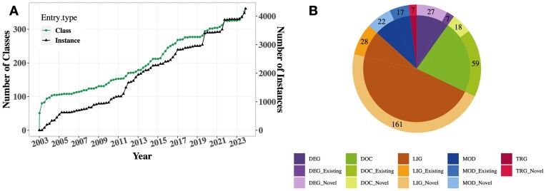
Figure 1.
Full legend
Data growth trends within the ELM resource since the last update paper (37). (A) This depicts the cumulative count of both motif classes and instances inserted into ELM in the last two decades. (B) The pie chart illustrates the allocation of new instances among both new and updated ELM classes (lighter and darker shades in the outer circle, respectively) belonging to 5 different functional categories. Note that the pie chart focuses only on the classes where new instances were added in this update. Visualisations were done using ggplot2 in RStudio (http://www.rstudio.com/).
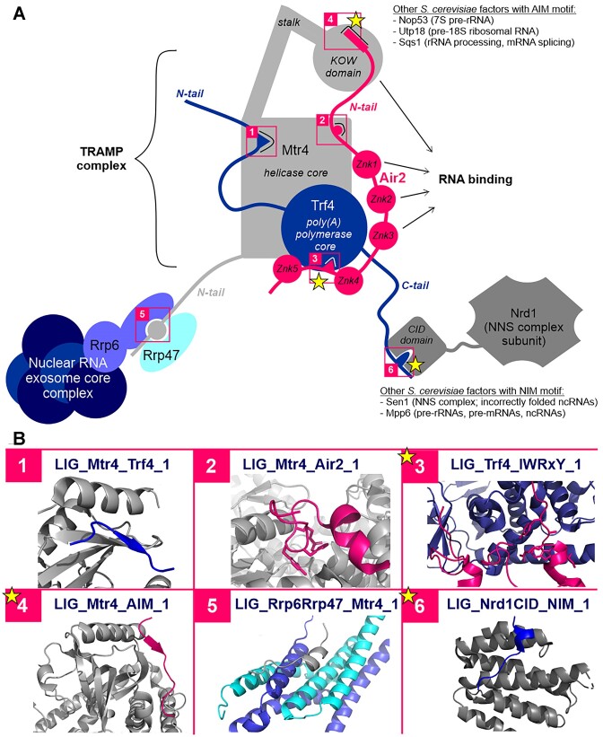
Figure 2.
Full legend
SLiMs in nuclear RNA processing and decay. (A) Schematic representation of the motif-mediated interactions maintaining the inter-subunit interactions of the TRAMP complex and facilitating the recruitment of the complex and the associated exosome to different types of degradable RNA. Motif-mediated interactions are highlighted with numbered pink boxes, with the ones newly added or extensively revised since the last release marked by a star. Where the given motif is employed by several yeast factors belonging to different protein families, the factors and their respective bound RNA types are listed. Abbreviations: CID, CTD-interacting domain, Znk, zinc knuckle, ncRNA - non-coding RNA, AIM, arch-interacting motif, NIM, Nrd1-interacting motif. (B) Structural snapshots of the numbered motif-mediated interactions. Colours for the domains and/or motifs depicted in each box match those of the corresponding proteins in the schematic figure of panel A. Within each box, all protein domains are depicted in cartoon representation. In most cases (boxes 1, 4, 5 and 6) cartoon representation was also applied to the SLiMs for simplicity. In the case of more extensive binding interfaces (boxes 2 and 3), where the motif residues would be difficult to guess, residues belonging to the SLiMs are shown in stick representation. The structural snapshots do not always show the interaction of the exact same proteins as depicted in panel A, in some cases, an X-ray structure was only available for the same type of motif from another protein. (1) PDB: 4U4C (Mtr4 with a Trf4-Air2 fusion protein, Trf4 motif depicted); (2) PDB:4U4C (Mtr4 with a Trf4-Air2 fusion protein, Air2 motif depicted); (3) PDB:3NYB (Trf4 with Air2 regions including the 4th and 5th zink knuckles and the motif in the linker connecting those; motif residues in stick representation); (4) PDB:5OOQ (Mtr4 KOW domain with the AIM motif from Nop53); (5) PDB:4WFD (the PMC2NT domain of Rrp6 forms a heterodimer with cofactor Rrp47 and the resulting dimer interface binds the motif within the N-terminal tail of Mtr4); (6) PDB: 6O3W (CID domain of Nrd1 bound by a Sen1 NIM motif).
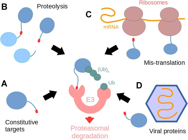
Figure 3.
Full legend
The diverse functions of C-terminal degrons. These motifs (red) can be part of native, endogenous protein C-termini (A), or be at an internal location and exposed by proteolysis (B). Degron SLiMs may also originate from erroneously translated mRNA sequences (C) or be present in virus-encoded proteins (D). These degrons are subsequently recognized by specific ubiquitin ligases (E3) and the proteins are polyubiquitylated, targeting them into the proteasome.
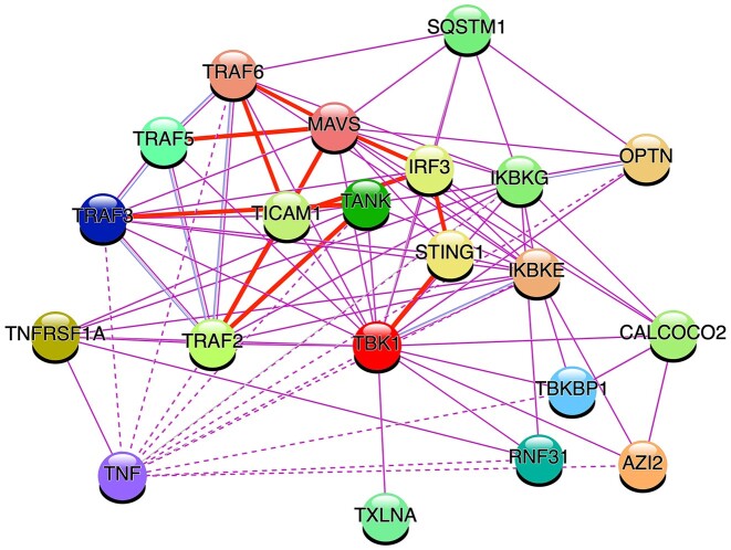
Figure 4.
Full legend
Protein interaction network centred on the key anti-pathogen and inflammatory kinase TBK1 generated by the STRING resource (122). The network shows how TBK1 plays a core role in bringing together the TRAF and IRF3 signalling systems. Edges with SLiMs annotated in ELM are shown by thick red lines. TNF (extracellular) interactions with cytosolic proteins are indirect (dotted lines). STRING settings for building the interaction network were as follows: only experimentally identified interactors of TBK1 and the 1st shell of ‘no more than 20 interactors’ in max number of interactors.
References122
- 1.
Pawson T., Nash P. Assembly of cell regulatory systems through protein interaction domains. Science. 2003; 300:445–452.
Not in our library10.1126/science.1083653 (opens in a new tab)
- 2.
Gibson T.J. Cell regulation: determined to signal discrete cooperation. Trends Biochem. Sci.2009; 34:471–482.
Not in our library10.1016/j.tibs.2009.06.007 (opens in a new tab)
- 3.
Wright P.E., Dyson H.J. Intrinsically disordered proteins in cellular signalling and regulation. Nat. Rev. Mol. Cell Biol.2015; 16:18–29.
Not in our library10.1038/nrm3920 (opens in a new tab)
- 4.
- 5.
Bah A., Forman-Kay J.D. Modulation of intrinsically disordered protein function by post-translational modifications. J. Biol. Chem.2016; 291:6696–6705.
Not in our library10.1074/jbc.r115.695056 (opens in a new tab)
- 6.
Pajkos M., Dosztányi Z. Functions of intrinsically disordered proteins through evolutionary lenses. Prog. Mol. Biol. Transl. Sci.2021; 183:45–74.
Not in our library10.1016/bs.pmbts.2021.06.017 (opens in a new tab)
- 7.
Van Roey K., Uyar B., Weatheritt R.J., Dinkel H., Seiler M., Budd A., Gibson T.J., Davey N.E. Short linear motifs: ubiquitous and functionally diverse protein interaction modules directing cell regulation. Chem. Rev.2014; 114:6733–6778.
Not in our library10.1021/cr400585q (opens in a new tab)
- 8.
Zanzoni A., Ribeiro D.M., Brun C. Understanding protein multifunctionality: from short linear motifs to cellular functions. Cell. Mol. Life Sci.2019; 76:4407–4412.
Not in our library10.1007/s00018-019-03273-4 (opens in a new tab)
- 9.
Kliche J., Ivarsson Y. Orchestrating serine/threonine phosphorylation and elucidating downstream effects by short linear motifs. Biochem. J.2022; 479:1–22.
Not in our library10.1042/bcj20200714 (opens in a new tab)
- 10.
Weikl T.R., Paul F. Conformational selection in protein binding and function. Protein Sci.2014; 23:1508–1518.
Not in our library10.1002/pro.2539 (opens in a new tab)
- 11.
- 12.
Tompa P., Davey N.E., Gibson T.J., Babu M.M. A million peptide motifs for the molecular biologist. Mol. Cell. 2014; 55:161–169.
Not in our library10.1016/j.molcel.2014.05.032 (opens in a new tab)
- 13.
Hunt T. Protein sequence motifs involved in recognition and targeting: a new series. Trends Biochem. Sci. 1990; 15:305.2204156
Not in our library
- 14.
Munro S., Pelham H.R. A C-terminal signal prevents secretion of luminal ER proteins. Cell. 1987; 48:899–907.
Not in our library10.1016/0092-8674(87)90086-9 (opens in a new tab)
- 15.
Dingwall C., Laskey R.A. Nuclear targeting sequences–a consensus?. Trends Biochem. Sci.1991; 16:478–481.
Not in our library10.1016/0968-0004(91)90184-w (opens in a new tab)
- 16.
Kalderon D., Roberts B.L., Richardson W.D., Smith A.E. A short amino acid sequence able to specify nuclear location. Cell. 1984; 39:499–509.
Not in our library10.1016/0092-8674(84)90457-4 (opens in a new tab)
- 17.
Gould S.J., Keller G.A., Subramani S. Identification of peroxisomal targeting signals located at the carboxy terminus of four peroxisomal proteins. J. Cell Biol.1988; 107:897–905.
Not in our library10.1083/jcb.107.3.897 (opens in a new tab)
- 18.
Van Roey K., Gibson T.J., Davey N.E. Motif switches: decision-making in cell regulation. Curr. Opin. Struct. Biol.2012; 22:378–385.
Not in our library10.1016/j.sbi.2012.03.004 (opens in a new tab)
- 19.
Robinson M.S. Adaptable adaptors for coated vesicles. Trends Cell Biol.2004; 14:167–174.
Not in our library10.1016/j.tcb.2004.02.002 (opens in a new tab)
- 20.
Lingaraju M., Johnsen D., Schlundt A., Langer L.M., Basquin J., Sattler M., Heick Jensen T., Falk S., Conti E. The MTR4 helicase recruits nuclear adaptors of the human RNA exosome using distinct arch-interacting motifs. Nat. Commun.2019; 10:3393.
Not in our library10.1038/s41467-019-11339-x (opens in a new tab)
- 21.
Giangreco G., Malabarba M.G., Sigismund S. Specialised endocytic proteins regulate diverse internalisation mechanisms and signalling outputs in physiology and cancer. Biol. Cell. 2021; 113:165–182.
Not in our library10.1111/boc.202000129 (opens in a new tab)
- 22.
- 23.
Hossain M., Bhalla K., Stillman B. Multiple, short protein binding motifs in ORC1 and CDC6 control the initiation of DNA replication. Mol. Cell. 2021; 81:1951–1969.
Not in our library10.1016/j.molcel.2021.03.003 (opens in a new tab)
- 24.
González-Foutel N.S., Glavina J., Borcherds W.M., Safranchik M., Barrera-Vilarmau S., Sagar A., Estaña A., Barozet A., Garrone N.A., Fernandez-Ballester G. et al. Conformational buffering underlies functional selection in intrinsically disordered protein regions. Nat. Struct. Mol. Biol.2022; 29:781–790.
Not in our library10.1038/s41594-022-00811-w (opens in a new tab)
- 25.
Case L.B., Ditlev J.A., Rosen M.K. Regulation of transmembrane signaling by phase separation. Annu. Rev. Biophys.2019; 48:465–494.
Not in our library10.1146/annurev-biophys-052118-115534 (opens in a new tab)
- 26.
Zeng M., Chen X., Guan D., Xu J., Wu H., Tong P., Zhang M. Reconstituted postsynaptic density as a molecular platform for understanding synapse formation and plasticity. Cell. 2018; 174:1172–1187.
Not in our library10.1016/j.cell.2018.06.047 (opens in a new tab)
- 27.
Leung G.C., Murphy J.M., Briant D., Sicheri F. Characterization of kinase target phosphorylation consensus motifs using peptide SPOT arrays. Methods Mol. Biol.2009; 570:187–195.
Not in our library10.1007/978-1-60327-394-7_7 (opens in a new tab)
- 28.
- 29.
Münger K., Werness B.A., Dyson N., Phelps W.C., Harlow E., Howley P.M. Complex formation of human papillomavirus E7 proteins with the retinoblastoma tumor suppressor gene product. EMBO J.1989; 8:4099–4105.
Not in our library10.1002/j.1460-2075.1989.tb08594.x (opens in a new tab)
- 30.
Davey N.E., Travé G., Gibson T.J. How viruses hijack cell regulation. Trends Biochem. Sci.2011; 36:159–169.
Not in our library10.1016/j.tibs.2010.10.002 (opens in a new tab)
- 31.
Madhu P., Davey N.E., Ivarsson Y. How viral proteins bind short linear motifs and intrinsically disordered domains. Essays Biochem.2022; 66:935–944.
Not in our library10.1042/ebc20220047 (opens in a new tab)
- 32.
Ali M., Simonetti L., Ivarsson Y. Screening intrinsically disordered regions for short linear binding motifs. Methods Mol. Biol.2020; 2141:529–552.
Not in our library10.1007/978-1-0716-0524-0_27 (opens in a new tab)
- 33.
Vincentelli R., Luck K., Poirson J., Polanowska J., Abdat J., Blémont M., Turchetto J., Iv F., Ricquier K., Straub M.-L. et al. Quantifying domain-ligand affinities and specificities by high-throughput holdup assay. Nat. Methods. 2015; 12:787–793.
Not in our library10.1038/nmeth.3438 (opens in a new tab)
- 34.
- 35.
Bandyopadhyay S., Bhaduri S., Örd M., Davey N.E., Loog M., Pryciak P.M. Comprehensive Analysis of G1 Cyclin Docking Motif Sequences that Control CDK Regulatory Potency In Vivo. Curr. Biol.2020; 30:4454–4466.
Not in our library10.1016/j.cub.2020.08.099 (opens in a new tab)
- 36.
Davey N.E., Simonetti L., Ivarsson Y. The next wave of interactomics: mapping the SLiM-based interactions of the intrinsically disordered proteome. Curr. Opin. Struct. Biol.2023; 80:102593.
Not in our library10.1016/j.sbi.2023.102593 (opens in a new tab)
- 37.
- 38.
Gouw M., Alvarado-Valverde J., Čalyševa J., Diella F., Kumar M., Michael S., Van Roey K., Dinkel H., Gibson T.J. How to annotate and submit a short linear motif to the eukaryotic linear motif resource. Methods Mol. Biol.2020; 2141:73–102.
Not in our library10.1007/978-1-0716-0524-0_4 (opens in a new tab)
- 39.
Houseley J., LaCava J., Tollervey D. RNA-quality control by the exosome. Nat. Rev. Mol. Cell Biol.2006; 7:529–539.
Not in our library10.1038/nrm1964 (opens in a new tab)
- 40.
Falk S., Weir J.R., Hentschel J., Reichelt P., Bonneau F., Conti E. The molecular architecture of the TRAMP complex reveals the organization and interplay of its two catalytic activities. Mol. Cell. 2014; 55:856–867.
Not in our library10.1016/j.molcel.2014.07.020 (opens in a new tab)
- 41.
Thoms M., Thomson E., Baßler J., Gnädig M., Griesel S., Hurt E. The exosome is recruited to RNA substrates through specific adaptor proteins. Cell. 2015; 162:1029–1038.
Not in our library10.1016/j.cell.2015.07.060 (opens in a new tab)
- 42.
- 43.
Losh J.S., King A.K., Bakelar J., Taylor L., Loomis J., Rosenzweig J.A., Johnson S.J., van Hoof A. Interaction between the RNA-dependent ATPase and poly(A) polymerase subunits of the TRAMP complex is mediated by short peptides and important for snoRNA processing. Nucleic Acids Res.2015; 43:1848–1858.
Not in our library10.1093/nar/gkv005 (opens in a new tab)
- 44.
Falk S., Tants J.-N., Basquin J., Thoms M., Hurt E., Sattler M., Conti E. Structural insights into the interaction of the nuclear exosome helicase Mtr4 with the preribosomal protein Nop53. RNA. 2017; 23:1780–1787.
Not in our library10.1261/rna.062901.117 (opens in a new tab)
- 45.
Wang J., Chen J., Wu G., Zhang H., Du X., Chen S., Zhang L., Wang K., Fan J., Gao S. et al. NRDE2 negatively regulates exosome functions by inhibiting MTR4 recruitment and exosome interaction. Genes Dev.2019; 33:536–549.
Not in our library10.1101/gad.322602.118 (opens in a new tab)
- 46.
Hamill S., Wolin S.L., Reinisch K.M. Structure and function of the polymerase core of TRAMP, a RNA surveillance complex. Proc. Natl. Acad. Sci. U.S.A.2010; 107:15045–15050.
Not in our library10.1073/pnas.1003505107 (opens in a new tab)
- 47.
Fasken M.B., Leung S.W., Banerjee A., Kodani M.O., Chavez R., Bowman E.A., Purohit M.K., Rubinson M.E., Rubinson E.H., Corbett A.H. Air1 zinc knuckles 4 and 5 and a conserved IWRXY motif are critical for the function and integrity of the Trf4/5-Air1/2-Mtr4 polyadenylation (TRAMP) RNA quality control complex. J. Biol. Chem.2011; 286:37429–37445.
Not in our library10.1074/jbc.m111.271494 (opens in a new tab)
- 48.
Tudek A., Porrua O., Kabzinski T., Lidschreiber M., Kubicek K., Fortova A., Lacroute F., Vanacova S., Cramer P., Stefl R. et al. Molecular basis for coordinating transcription termination with noncoding RNA degradation. Mol. Cell. 2014; 55:467–481.
Not in our library10.1016/j.molcel.2014.05.031 (opens in a new tab)
- 49.
Zhang Y., Chun Y., Buratowski S., Tong L. Identification of three sequence motifs in the transcription termination factor Sen1 that mediate direct interactions with Nrd1. Structure. 2019; 27:1156–1161.
Not in our library10.1016/j.str.2019.04.005 (opens in a new tab)
- 50.
Kim K., Heo D.-H., Kim I., Suh J.-Y., Kim M. Exosome cofactors connect transcription termination to RNA processing by guiding terminated transcripts to the appropriate exonuclease within the nuclear exosome. J. Biol. Chem.2016; 291:13229–13242.
Not in our library10.1074/jbc.m116.715771 (opens in a new tab)
- 51.
YAMAMOTO M. The selective elimination of messenger RNA underlies the mitosis–meiosis switch in fission yeast. Proc. Jpn. Acad., Ser. B. 2010; 86:788–797.
Not in our library10.2183/pjab.86.788 (opens in a new tab)
- 52.
Egan E.D., Braun C.R., Gygi S.P., Moazed D. Post-transcriptional regulation of meiotic genes by a nuclear RNA silencing complex. RNA. 2014; 20:867–881.
Not in our library10.1261/rna.044479.114 (opens in a new tab)
- 53.
Zhou Y., Zhu J., Schermann G., Ohle C., Bendrin K., Sugioka-Sugiyama R., Sugiyama T., Fischer T. The fission yeast MTREC complex targets CUTs and unspliced pre-mRNAs to the nuclear exosome. Nat. Commun.2015; 6:7050.
Not in our library10.1038/ncomms8050 (opens in a new tab)
- 54.
Sugiyama T., Sugioka-Sugiyama R. Red1 promotes the elimination of meiosis-specific mRNAs in vegetatively growing fission yeast. EMBO J.2011; 30:1027–1039.
Not in our library10.1038/emboj.2011.32 (opens in a new tab)
- 55.
Schulze W.M., Stein F., Rettel M., Nanao M., Cusack S. Structural analysis of human ARS2 as a platform for co-transcriptional RNA sorting. Nat. Commun.2018; 9:1701.
Not in our library10.1038/s41467-018-04142-7 (opens in a new tab)
- 56.
- 57.
Rouvière J.O., Salerno-Kochan A., Lykke-Andersen S., Garland W., Dou Y., Rathore O., Molska E.Š., Wu G., Schmid M., Bugai A. et al. ARS2 instructs early transcription termination-coupled RNA decay by recruiting ZC3H4 to nascent transcripts. Mol. Cell. 2023; 83:2240–2257.
Not in our library10.1016/j.molcel.2023.05.028 (opens in a new tab)
- 58.
Yeh C.-W., Huang W.-C., Hsu P.-H., Yeh K.-H., Wang L.-C., Hsu P.W.-C., Lin H.-C., Chen Y.-N., Chen S.-C., Yeang C.-H. et al. The C-degron pathway eliminates mislocalized proteins and products of deubiquitinating enzymes. EMBO J.2021; 40:e105846.
Not in our library10.15252/embj.2020105846 (opens in a new tab)
- 59.
Varshavsky A. N-degron and C-degron pathways of protein degradation. P. Natl. Acad. Sci. U.S.A.2019; 116:358–366.
Not in our library10.1073/pnas.1816596116 (opens in a new tab)
- 60.
Chen X., Liao S., Makaros Y., Guo Q., Zhu Z., Krizelman R., Dahan K., Tu X., Yao X., Koren I. et al. Molecular basis for arginine C-terminal degron recognition by Cul2FEM1 E3 ligase. Nat. Chem. Biol.2021; 17:254–262.
Not in our library10.1038/s41589-020-00704-3 (opens in a new tab)
- 61.
Mészáros B., Kumar M., Gibson T.J., Uyar B., Dosztányi Z. Degrons in cancer. Sci. Signal.2017; 10:eaak9982.
Not in our library10.1126/scisignal.aak9982 (opens in a new tab)
- 62.
Timms R.T., Koren I. Tying up loose ends: the N-degron and C-degron pathways of protein degradation. Biochem. Soc. Trans.2020; 48:1557–1567.
Not in our library10.1042/bst20191094 (opens in a new tab)
- 63.
Koren I., Timms R.T., Kula T., Xu Q., Li M.Z., Elledge S.J. The Eukaryotic Proteome Is Shaped by E3 Ubiquitin Ligases Targeting C-Terminal Degrons. Cell. 2018; 173:1622–1635.
Not in our library10.1016/j.cell.2018.04.028 (opens in a new tab)
- 64.
Sitron C.S., Brandman O. CAT tails drive degradation of stalled polypeptides on and off the ribosome. Nat. Struct. Mol. Biol.2019; 26:450–459.
Not in our library10.1038/s41594-019-0230-1 (opens in a new tab)
- 65.
Rusnac D.-V., Lin H.-C., Canzani D., Tien K.X., Hinds T.R., Tsue A.F., Bush M.F., Yen H.-C.S., Zheng N. Recognition of the diglycine C-end degron by CRL2KLHDC2 ubiquitin ligase. Mol. Cell. 2018; 72:813–822.
Not in our library10.1016/j.molcel.2018.10.021 (opens in a new tab)
- 66.
Okumura F., Fujiki Y., Oki N., Osaki K., Nishikimi A., Fukui Y., Nakatsukasa K., Kamura T. Cul5-type ubiquitin ligase KLHDC1 contributes to the elimination of truncated SELENOS produced by failed UGA/Sec decoding. iScience. 2020; 23:100970.
Not in our library10.1016/j.isci.2020.100970 (opens in a new tab)
- 67.
Ru Y., Yan X., Zhang B., Song L., Feng Q., Ye C., Zhou Z., Yang Z., Li Y., Zhang Z. et al. C-terminal glutamine acts as a C-degron targeted by E3 ubiquitin ligase TRIM7. Proc. Natl. Acad. Sci. U.S.A.2022; 119:e2203218119.
Not in our library10.1073/pnas.2203218119 (opens in a new tab)
- 68.
Pla-Prats C., Cavadini S., Kempf G., Thomä N.H. Recognition of the CCT5 di-Glu degron by CRL4DCAF12 is dependent on TRiC assembly. EMBO J.2023; 42:e112253.
Not in our library10.15252/embj.2022112253 (opens in a new tab)
- 69.
Ravichandran R., Kodali K., Peng J., Potts P.R. Regulation of MAGE-A3/6 by the CRL4-DCAF12 ubiquitin ligase and nutrient availability. EMBO Rep.2019; 20:e47352.
Not in our library10.15252/embr.201847352 (opens in a new tab)
- 70.
Jiao D., Chen Y., Wang Y., Sun H., Shi Q., Zhang L., Zhao X., Liu Y., He H., Lv Z. et al. DCAF12 promotes apoptosis and inhibits NF-κB activation by acting as an endogenous antagonist of IAPs. Oncogene. 2022; 41:3000–3010.
Not in our library10.1038/s41388-022-02319-5 (opens in a new tab)
- 71.
Lidak T., Baloghova N., Korinek V., Sedlacek R., Balounova J., Kasparek P., Cermak L. CRL4-DCAF12 ubiquitin ligase controls MOV10 RNA helicase during spermatogenesis and T cell activation. Int. J. Mol. Sci.2021; 22:5394.
Not in our library10.3390/ijms22105394 (opens in a new tab)
- 72.
Timms R.T., Mena E.L., Leng Y., Li M.Z., Tchasovnikarova I.A., Koren I., Elledge S.J. Defining E3 ligase-substrate relationships through multiplex CRISPR screening. Nat. Cell Biol.2023; 25:1535–1545.
Not in our library10.1038/s41556-023-01229-2 (opens in a new tab)
- 73.
Scott D.C., King M.T., Baek K., Gee C.T., Kalathur R., Li J., Purser N., Nourse A., Chai S.C., Vaithiyalingam S. et al. E3 ligase autoinhibition by C-degron mimicry maintains C-degron substrate fidelity. Mol. Cell. 2023; 83:770–786.
Not in our library10.1016/j.molcel.2023.01.019 (opens in a new tab)
- 74.
Lin H.-C., Ho S.-C., Chen Y.-Y., Khoo K.-H., Hsu P.-H., Yen H.-C.S. SELENOPROTEINS. CRL2 aids elimination of truncated selenoproteins produced by failed UGA/Sec decoding. Science. 2015; 349:91–95.
Not in our library10.1126/science.aab0515 (opens in a new tab)
- 75.
Hwang J., Qi L. Quality control in the endoplasmic reticulum: crosstalk between ERAD and UPR pathways. Trends Biochem. Sci.2018; 43:593–605.
Not in our library10.1016/j.tibs.2018.06.005 (opens in a new tab)
- 76.
Guerriero C.J., Brodsky J.L. The delicate balance between secreted protein folding and endoplasmic reticulum-associated degradation in human physiology. Physiol. Rev.2012; 92:537–576.
Not in our library10.1152/physrev.00027.2011 (opens in a new tab)
- 77.
Zhang J., Lan Y., Sanyal S. Membrane heist: coronavirus host membrane remodeling during replication. Biochimie. 2020; 179:229–236.
Not in our library10.1016/j.biochi.2020.10.010 (opens in a new tab)
- 78.
Noack J., Bernasconi R., Molinari M. How viruses hijack the ERAD tuning machinery. J. Virol.2014; 88:10272–10275.
Not in our library10.1128/jvi.00801-14 (opens in a new tab)
- 79.
- 80.
Schaeffer V., Akutsu M., Olma M.H., Gomes L.C., Kawasaki M., Dikic I. Binding of OTULIN to the PUB domain of HOIP controls NF-κB signaling. Mol. Cell. 2014; 54:349–361.
Not in our library10.1016/j.molcel.2014.03.016 (opens in a new tab)
- 81.
Conner S.D., Schmid S.L. Identification of an adaptor-associated kinase, AAK1, as a regulator of clathrin-mediated endocytosis. J. Cell Biol.2002; 156:921–929.
Not in our library10.1083/jcb.200108123 (opens in a new tab)
- 82.
Karim M., Saul S., Ghita L., Sahoo M.K., Ye C., Bhalla N., Lo C.-W., Jin J., Park J.-G., Martinez-Gualda B. et al. Numb-associated kinases are required for SARS-CoV-2 infection and are cellular targets for antiviral strategies. Antiviral Res.2022; 204:105367.
Not in our library10.1016/j.antiviral.2022.105367 (opens in a new tab)
- 83.
- 84.
Pu S., Schor S., Karim M., Saul S., Robinson M., Kumar S., Prugar L.I., Dorosky D.E., Brannan J., Dye J.M. et al. BIKE regulates dengue virus infection and is a cellular target for broad-spectrum antivirals. Antiviral Res.2020; 184:104966.
Not in our library10.1016/j.antiviral.2020.104966 (opens in a new tab)
- 85.
Sorensen E.B., Conner S.D. AAK1 regulates Numb function at an early step in clathrin-mediated endocytosis. Traffic. 2008; 9:1791–1800.
Not in our library10.1111/j.1600-0854.2008.00790.x (opens in a new tab)
- 86.
Ricotta D., Conner S.D., Schmid S.L., von Figura K., Honing S. Phosphorylation of the AP2 mu subunit by AAK1 mediates high affinity binding to membrane protein sorting signals. J. Cell Biol.2002; 156:791–795.
Not in our library10.1083/jcb.200111068 (opens in a new tab)
- 87.
Liu Q., Bautista-Gomez J., Higgins D.A., Yu J., Xiong Y. Dysregulation of the AP2M1 phosphorylation cycle by LRRK2 impairs endocytosis and leads to dopaminergic neurodegeneration. Sci. Signal.2021; 14:eabg3555.
Not in our library10.1126/scisignal.abg3555 (opens in a new tab)
- 88.
Zeng G., Cai M. Regulation of the actin cytoskeleton organization in yeast by a novel serine/threonine kinase Prk1p. J. Cell Biol.1999; 144:71–82.
Not in our library10.1083/jcb.144.1.71 (opens in a new tab)
- 89.
Zeng G., Yu X., Cai M. Regulation of yeast actin cytoskeleton-regulatory complex Pan1p/Sla1p/End3p by serine/threonine kinase Prk1p. Mol. Biol. Cell. 2001; 12:3759–3772.
Not in our library10.1091/mbc.12.12.3759 (opens in a new tab)
- 90.
Henry K.R., D’Hondt K., Chang J.S., Nix D.A., Cope M.J.T.V., Chan C.S.M., Drubin D.G., Lemmon S.K The actin-regulating kinase Prk1p negatively regulates Scd5p, a suppressor of clathrin deficiency, in actin organization and endocytosis. Curr. Biol.2003; 13:1564–1569.
Not in our library10.1016/s0960-9822(03)00579-7 (opens in a new tab)
- 91.
Belkina N.V., Liu Y., Hao J.-J., Karasuyama H., Shaw S. LOK is a major ERM kinase in resting lymphocytes and regulates cytoskeletal rearrangement through ERM phosphorylation. Proc. Natl. Acad. Sci. U.S.A.2009; 106:4707–4712.
Not in our library10.1073/pnas.0805963106 (opens in a new tab)
- 92.
Shah N.H., Amacher J.F., Nocka L.M., Kuriyan J. The Src module: an ancient scaffold in the evolution of cytoplasmic tyrosine kinases. Crit. Rev. Biochem. Mol. Biol.2018; 53:535–563.
Not in our library10.1080/10409238.2018.1495173 (opens in a new tab)
- 93.
Buday L., Wunderlich L., Tamás P. The Nck family of adapter proteins: regulators of actin cytoskeleton. Cell. Signal.2002; 14:723–731.
Not in our library10.1016/s0898-6568(02)00027-x (opens in a new tab)
- 94.
Huang H., Li L., Wu C., Schibli D., Colwill K., Ma S., Li C., Roy P., Ho K., Songyang Z. et al. Defining the specificity space of the human SRC homology 2 domain. Mol. Cell. Proteomics. 2008; 7:768–784.
Not in our library10.1074/mcp.m700312-mcp200 (opens in a new tab)
- 95.
Sámano-Sánchez H., Gibson T.J., Chemes L.B. Using linear motif database resources to identify SH2 domain binders. Methods Mol. Biol.2023; 2705:153–197.
Not in our library10.1007/978-1-0716-3393-9_9 (opens in a new tab)
- 96.
Frese S., Schubert W.-D., Findeis A.C., Marquardt T., Roske Y.S., Stradal T.E.B., Heinz D.W. The phosphotyrosine peptide binding specificity of Nck1 and Nck2 Src homology 2 domains. J. Biol. Chem.2006; 281:18236–18245.
Not in our library10.1074/jbc.m512917200 (opens in a new tab)
- 97.
- 98.
Zevini A., Olagnier D., Hiscott J. Crosstalk between cytoplasmic RIG-I and STING sensing pathways. Trends Immunol.2017; 38:194–205.
Not in our library10.1016/j.it.2016.12.004 (opens in a new tab)
- 99.
Chen Q., Sun L., Chen Z.J. Regulation and function of the cGAS-STING pathway of cytosolic DNA sensing. Nat. Immunol.2016; 17:1142–1149.
Not in our library10.1038/ni.3558 (opens in a new tab)
- 100.
Hildebrand J.M., Yi Z., Buchta C.M., Poovassery J., Stunz L.L., Bishop G.A. Roles of tumor necrosis factor receptor associated factor 3 (TRAF3) and TRAF5 in immune cell functions. Immunol. Rev.2011; 244:55–74.
Not in our library10.1111/j.1600-065x.2011.01055.x (opens in a new tab)
- 101.
Bishop G.A. TRAF3 as a powerful and multitalented regulator of lymphocyte functions. J. Leukoc. Biol.2016; 100:919–926.
Not in our library10.1189/jlb.2mr0216-063r (opens in a new tab)
- 102.
Liu B., Gao C. Regulation of MAVS activation through post-translational modifications. Curr. Opin. Immunol.2018; 50:75–81.
Not in our library10.1016/j.coi.2017.12.002 (opens in a new tab)
- 103.
Ortega E., Rengachari S., Ibrahim Z., Hoghoughi N., Gaucher J., Holehouse A.S., Khochbin S., Panne D. Transcription factor dimerization activates the p300 acetyltransferase. Nature. 2018; 562:538–544.
Not in our library10.1038/s41586-018-0621-1 (opens in a new tab)
- 104.
Zhao B., Du F., Xu P., Shu C., Sankaran B., Bell S.L., Liu M., Lei Y., Gao X., Fu X. et al. A conserved PLPLRT/SD motif of STING mediates the recruitment and activation of TBK1. Nature. 2019; 569:718–722.
Not in our library10.1038/s41586-019-1228-x (opens in a new tab)
- 105.
Al Hamrashdi M., Brady G. Regulation of IRF3 activation in human antiviral signaling pathways. Biochem. Pharmacol.2022; 200:115026.
Not in our library10.1016/j.bcp.2022.115026 (opens in a new tab)
- 106.
Guharoy M., Lazar T., Macossay-Castillo M., Tompa P. Degron masking outlines degronons, co-degrading functional modules in the proteome. Commun. Biol.2022; 5:445.
Not in our library10.1038/s42003-022-03391-z (opens in a new tab)
- 107.
- 108.
- 109.
- 110.
Tusnády G.E., Zeke A., Kalman Z.E., Fatoux M., Ricard-Blum S., Gibson T.J., Dobson L. LeishMANIAdb: a comparative resource for Leishmania proteins. Database. 2023; 2023:baad074.
Not in our library10.1093/database/baad074 (opens in a new tab)
- 111.
Bret H., Andreani J., Guerois R. From interaction networks to interfaces: Scanning intrinsically disordered regions using AlphaFold2. 2023; bioRxiv doi:25 May 2023, preprint: not peer reviewed.
Not in our library10.1101/2023.05.25.542287 (opens in a new tab)
- 112.
Lee C.Y., Hubrich D., Varga J.K., Schäfer C., Welzel M., Schumbera E., Đokić M., Strom J.M., Schönfeld J., Geist J.L. et al. Systematic discovery of protein interaction interfaces using AlphaFold and experimental validation. 2023; bioRxiv doi:09 August 2023, preprint: not peer reviewed.
Not in our library10.1101/2023.08.07.552219 (opens in a new tab)
- 113.
Diella F., Haslam N., Chica C., Budd A., Michael S., Brown N.P., Trave G., Gibson T.J. Understanding eukaryotic linear motifs and their role in cell signaling and regulation. Front. Biosci.2008; 13:6580–6603.
Not in our library10.2741/3175 (opens in a new tab)
- 114.
Van Roey K., Davey N.E. Motif co-regulation and co-operativity are common mechanisms in transcriptional, post-transcriptional and post-translational regulation. Cell Commun. Signal.2015; 13:45.
Not in our library10.1186/s12964-015-0123-9 (opens in a new tab)
- 115.
Uyar B., Weatheritt R.J., Dinkel H., Davey N.E., Gibson T.J. Proteome-wide analysis of human disease mutations in short linear motifs: neglected players in cancer?. Mol. Biosyst.2014; 10:2626–2642.
Not in our library10.1039/c4mb00290c (opens in a new tab)
- 116.
Mészáros B., Sámano-Sánchez H., Alvarado-Valverde J., Čalyševa J., Martínez-Pérez E., Alves R., Shields D.C., Kumar M., Rippmann F., Chemes L.B. et al. Short linear motif candidates in the cell entry system used by SARS-CoV-2 and their potential therapeutic implications. Sci. Signal.2021; 14:eabd0334.
Not in our library10.1126/scisignal.abd0334 (opens in a new tab)
- 117.
Pittner N.A., Solomon R.N., Bui D.-C., McBride J.W. Ehrlichia effector SLiM-icry: artifice of cellular subversion. Front. Cell. Infect. Microbiol.2023; 13:1150758.
Not in our library10.3389/fcimb.2023.1150758 (opens in a new tab)
- 118.
Goswami S., Samanta D., Duraivelan K. Molecular mimicry of host short linear motif-mediated interactions utilised by viruses for entry. Mol. Biol. Rep.2023; 50:4665–4673.
Not in our library10.1007/s11033-023-08389-2 (opens in a new tab)
- 119.
- 120.
- 121.
InterPro in 2022 · 2022
- 122.
Reads
Read by people on this page
None yet.
Checked or read by AI through naulon
None yet.
A person counts once a day per paper. We keep no cookie and no address: a reader is a hash that changes every day. Any month under five reads shows no number.
Are you an author?
Is this your paper?
Claim it to add your note and be paid when agents cite it. Free.