Interaction between growing oocytes and granulosa cells in vitro
Md Hasanur Alam, Takashi Miyano
Reproductive Medicine and Biology · 2019 · Review · CC BY
doi:10.1002/rmb2.12292 (opens in a new tab)
We email you when a new notice or a question about this paper appears.
Abstract
Background. Oocyte growth is accompanied by follicular development in mammalian ovaries. Since the discovery of two oocyte‐derived factors, growth differentiation factor 9 (GDF9), and bone morphogenetic protein 15 (BMP15), knowledge of the bidirectional communication between oocytes and granulosa cells for ovarian function and fertility has been accumulated. In addition, the growth culture system of oocytes has been improved, further promoting the studies on the communication between oocytes and granulosa cells in vitro.
Methods. We provide an overview of the role of granulosa cells in oocyte growth and the role of oocytes in follicular development along with our recent findings in culture experiments of bovine growing oocytes.
Main findings. Granulosa cells supply nutrients and metabolites through gap junctions to oocytes and secrete paracrine signals to regulate oocytes. Oocytes regulate granulosa cell proliferation and differentiation and induce antrum formation via GDF9 and BMP15.
Conclusion. Oocytes actively participate in various aspects of follicular development, including antrum formation via the oocyte‐derived factors GDF9 and BMP15, whose synthesis is probably regulated by granulosa cells. In vitro studies will reveal the precise communication loop between oocytes and granulosa cells that facilitates the coordinated development of oocytes and granulosa cells in the follicles.
Abstract licence: CC BY
Full text
From PubMed Central, U.S. National Library of Medicine, as current on Oct 21, 2026. Paragraph numbers are ours, so an answer can point at one.
INTRODUCTION
Mammalian oogenesis starts at the embryonic period, and primordial germ cells (PGCs) are the primary cells in the process (Figure 1). After migration to the embryonic gonads, PGCs become oogonia and proliferate by mitosis. The oogonia subsequently enter meiosis I, at which point they are called oocytes, and become arrested at the diplotene stage of meiosis I.1 Individual oocytes become enclosed by a single layer of flattened pre‐granulosa cells in primordial follicles.2 When the oocytes start to grow, the surrounding pre‐granulosa cells become cubic granulosa cells in the primary follicles.3 The granulosa cells proliferate and form a multilayered structure, which is then further surrounded by layers of theca cells. Follicles at this stage are called secondary follicles. Later, a fluid‐filled cavity is formed inside the follicles and they become antral follicles. At this stage, the granulosa cells differentiate to cumulus granulosa cells which enclose the oocytes, and mural granulosa cells which form the inner layer of the follicle wall.4 In the meantime, the oocytes increase in size and prepare themselves for future maturation and fertilization with spermatozoa. After female animals reach puberty, the periodic gonadotropic surge induces fully grown oocytes in the antral follicles to resume meiosis I, to mature to metaphase II (MII), and finally to be ovulated.
The development of follicles is regulated by the hormones synthesized at the different levels in the hypothalamic‐pituitary‐ovarian axis: The hypothalamus secretes gonadotropin‐releasing hormone, the anterior pituitary secretes follicle‐stimulating hormone (FSH) and luteinizing hormone (LH), and the ovary produces steroidal hormones.5 In the follicles, FSH stimulates granulosa cell proliferation and aromatization of androgens to estrogens. Estrogens also stimulate granulosa cell proliferation.6 Previous studies have demonstrated that FSH receptors are expressed in the follicles from the primary to later stages 7 and that treatment with FSH promotes development of preantral follicles.8 Other studies have analyzed the growth‐promoting 6 and anti‐apoptotic actions 9 of FSH in the antral follicles. It has long been known that FSH is the predominant regulator of follicular development.
In addition to the endocrinological studies, ultrastructural studies have revealed the presence of direct connections between oocytes and granulosa cells. Anderson and Albertini showed the presence of heterologous gap junctions between oocytes and granulosa cells.10 Further, demonstration of lucifer yellow dye transfer from oocytes to granulosa cells clearly showed that there is direct communication through the gap junctions.11 Through the gap junctions, granulosa cells efficiently provide small molecules, such as nutrients, metabolic precursors, and molecular signals that regulate the oocytes.12 Therefore, prior to the discovery of the oocyte‐derived growth factors, it was thought that oocyte growth was controlled unidirectionally, with the surrounding granulosa cells supporting oocytes nutritionally, and FSH regulating the proliferation of granulosa cells.
In 1977, Eppig performed the first systematic study of in vitro growth (IVG) culture of oocytes,13 and it has since undergone continuous development as a new technology for utilizing incompetent oocytes in the ovary as a source of mature oocytes (Figure 1). In mice, application of IVG culture was extended from growing oocytes in secondary follicles 13 to non‐growing oocytes in primordial follicles in 1996,14 and to the production of baby mice from oogonia in fetal gonads in 2016.15 Further, Saitou and his colleagues produced PGC‐like cells from embryonic stem cells (ES cells) and induced pluripotent stem cells (iPS cells) derived from embryonic fibroblasts and adult tail tip fibroblasts,16, 17 and Hayashi and his colleagues generated fully potent mature oocytes completely in culture from ES cells and iPS cells.18 Our group has focused on IVG culture of domestic species, which would provide a new source of mature eggs for livestock production by using existing assisted reproductive technologies, such as in vitro maturation (IVM) and in vitro fertilization (IVF) of oocytes. Although progress on these technologies is far behind that of their counterparts in mice, the culture systems have been improved, and now, bovine growing oocytes collected from early antral follicles are able to grow to their final size and acquire the full developmental capacity efficiently.19 In both mice and domestic species, oocyte‐granulosa cell complexes are cultured, and most researchers have tried to maintain the granulosa cell viability and oocyte‐granulosa cell attachment during the long‐term culture period, because direct association of surrounding granulosa cells with oocytes through gap junctions is crucial for the oocyte viability and growth in vitro.
In addition to the endocrinological control of follicular development and the support provided to oocytes by granulosa cells, a new group of players joined the field of follicular development about two decades ago. These are the oocyte‐derived growth factors: growth differentiation factor 9 (GDF9) and bone morphogenetic protein 15 (BMP15).20, 21 Oocytes carry on their conversation with these factors inside the follicles, and follicular development is under the bidirectional communication between oocytes and granulosa cells.22 Now, a bovine IVG system for growing oocytes is nearly established.23, 24 Just as IVF in different species has provided a new understanding of mammalian fertilization and IVM has elucidated many aspects of the molecular control mechanisms of oocyte maturation, IVG of oocytes is expected to provide a new understanding of the mechanisms regulating the complex process of follicular development and oocyte growth in the mammalian ovary.
In this review, the bidirectional communication between oocytes and granulosa cells is outlined. We briefly review the role of granulosa cells in oocyte growth and the role of oocytes in follicular development, and reveal a new role of oocytes in antrum formation via oocyte‐derived growth factors based on our recent findings in bovine IVG experiments. For an explanation of the IVG culture systems used in our study, we recommend the reviews of Hirao.23, 24, 25
Structural connection between oocytes and granulosa cells
In primordial follicles, non‐growing oocytes are directly adjacent to surrounding pre‐granulosa cells. Shortly after oocytes enter the growth phase, an extracellular coat called the zona pellucida is assembled around the oocytes. Even after zona pellucida formation, however, the granulosa cells maintain contact with the oocytes via cytoplasmic processes known as transzonal projections (TZPs), which penetrate the zona pellucida (Figure 2). TZPs originate from granulosa cells, and some of them terminate at the oolemma to provide a means of direct connection between oocytes and granulosa cells.10, 26, 27 Most TZPs are composed of a strong backbone made of actin filaments,28 whereas a much smaller number of TZPs contain tubulin.29 Multiple TZPs typically project from each granulosa cell adjacent to the zona pellucida, while long actin‐rich filaments also project from some granulosa cells located in layers more distal to the oocyte.27, 30, 31 A dynamic change in the number and shape of TZPs occurs during follicular development. In growing oocytes, numerous TZPs develop and contribute to the growth. However, during the maturation of fully grown oocytes following the gonadotropic surge, active retraction of TZPs has been noted.32 In IVG of growing oocytes from domestic species, the number of TZPs significantly decreased during the culture, although the decrease was prevented by estradiol 17β for bovine oocytes,33 and by FSH for porcine oocytes.34
At the tip of TZPs, granulosa cells form heterologous gap junctions with oocytes. Gap junctions are intercellular channels that permit the direct transfer of ions and small molecules (<1 kDa) between adjacent cells.35 Gap junction channels are composed of connexins (Cx), a family of more than 20 members.36 Six connexins oligomerize to form a connexon (gap junction hemichannel), and two connexons in adjacent cells (between oocyte and granulosa cells, and between granulosa cell and granulosa cell in the follicle) dock to make a channel between the cells.37 Ovarian follicles of rodents express Cx32, Cx37, Cx43, and Cx45.38 Cx43 expression is restricted to the granulosa cells, whereas Cx37 is expressed exclusively in the oocytes.39 Bovine follicles express Cx26, Cx32, Cx37, and Cx43 40, 41; Cx43 is localized in granulosa cells 41, 42; and Cx26 is detected in oocytes.40 In the bovine follicles, Cx37 is expressed in both the oocytes and granulosa cells.42
Gap junctional channels transfer lucifer yellow dye, radiolabeled uridine metabolites, and electrical current in hamster oocyte‐granulosa cell complexes.11 Other studies suggested that granulosa cells provide nutrients, metabolic precursors, and signaling molecules through gap junctional channels to the oocytes.12, 26 Moreover, granulosa cells are coupled together via homologous gap junctions, so that the whole follicle including the oocyte, but not the theca cells, which are separated by the basement membrane, makes a functional syncytium.43 Small molecules such as amino acids, nucleotides, metabolites, and cyclic adenosine 3′,5′‐monophosphate (cAMP) are also exchanged through gap junctions among granulosa cells. Mice lacking Cx37 never develop mature Graafian follicles.44, 45 Cx37 deletion causes oocytes to arrest their growth at 74% in diameter of normal size and fail to achieve full meiotic competence.45
Amino acids and energy substrates
Through heterologous gap junctions, granulosa cells transport nutrients such as amino acids 46, 47 and substrates for energy production 48 to the oocytes. Six amino acid transport systems have been identified in mouse growing oocytes.49 Although the presence of granulosa cells surrounding growing oocytes does not confer amino acid transport by additional transport systems not present in the oocytes, the granulosa cells enhance the uptake of glycine, alanine, lysine, and taurine by oocytes, perhaps via gap junctions.49
Glucose uptake by oocytes occurs via the facilitative glucose transporter (GLUT) proteins in mice,50 cows,51 sheep,52 humans,53 and rhesus monkeys.54 But mammalian oocytes have low capacity to utilize glucose as a substrate,50, 55, 56, 57, 58, 59 possibly due to having a limited amount of the glycolytic enzyme phosphofructokinase.60 Thus, oocytes in most mammalian species appear to rely on granulosa cells that contain an additional GLUT with high affinity to glucose and high phosphofructokinase activity to convert glucose into readily utilized substrates (ie, pyruvate, lactate, NADPH etc).55, 58 These substrates are used by oocytes for the energy metabolism necessary for oocyte growth.59 Oocytes denuded of their granulosa cells are able to utilize pyruvate and other intermediaries of the tricarboxylic acid pathway for energy production, but not glucose.60
Granulosa cell–free culture systems have been reported in which naked mouse oocytes grew to 35 µm in diameter and formed the zona pellucida, and some of the oocytes reached around 70 µm in the existence of cocultured thecal stem cells.61 However, in general, the rate of oocyte growth in vitro is directly correlated with the number of granulosa cells coupled to a given oocyte.26 In domestic species, growing oocytes normally become degenerated when they detach from granulosa cells in IVG culture.25 Direct association with granulosa cells supports oocyte viability and growth through the gap junctions, which serve as efficient passages for amino acids and energy substrates.
cAMP and cGMP
During the growth phase, oocytes are arrested at the prophase of meiosis I. After reaching their full size, oocytes in the large antral follicles resume meiosis in response to the FSH + LH surge. Small oocytes in the primordial, primary, and secondary follicles (even in the early antral follicles in large mammals) have no ability to resume meiosis.62 During the final growth phase, oocytes acquire meiotic competence in a stepwise manner; first, they acquire the competence to resume meiosis and then become competent to progress to MII.63 However, after oocytes become competent, they do not resume meiosis in the follicle until being stimulated by the gonadotropic surge. Meiotic resumption of oocytes is prevented by the inhibitory influence of the follicular environment, mainly by inhibitory substances produced by granulosa cells. Thus, after such competent oocytes are released from the follicle environment, they resume meiosis spontaneously without gonadotrophic hormones,64 especially after denudation of the surrounding granulosa cells.65
Spontaneous meiotic resumption of isolated oocytes is proceeded by a drop in intracellular levels of cAMP.66 Several studies have suggested that cAMP derived from cumulus granulosa cells maintains the meiotic arrest of oocytes 67, 68 (Figure 2). Another cyclic nucleotide, cyclic guanosine 3′,5′‐monophosphate (cGMP), also plays a role in the maintenance of oocyte meiotic arrest. cGMP passes through gap junctions into the oocytes, where it inhibits phosphodiesterase 3 (PDE3), a hydrolytic enzyme of cAMP. It has been well established in mice that the inhibition of PDE3 maintains a high concentration of cAMP in the oocytes in order to block the meiotic resumption.69 These cyclic nucleotides act as negative regulators of the meiotic resumption of oocytes.
cAMP is synthesized by adenylyl cyclase, and cGMP is synthesized by guanylyl cyclase. cAMP is degraded to 5′‐AMP, and cGMP is degraded to 5′‐GMP by a group of enzymes known as phosphodiesterases (PDEs). Earlier experiments to assess the effects of inhibiting PDE activity on the meiotic resumption of cumulus‐enclosed and denuded, fully grown oocytes were performed with 3‐isobutyl‐1‐methylxanthine (IBMX), a non‐specific inhibitor of PDEs. This inhibitor prevented the meiotic resumption of oocytes in different species, including rodents 70 and cows.71 Specific inhibition of PDE3 family members, but not of PDE4 isoforms, prevented the spontaneous maturation of rat,72 mouse,73 and porcine 74 oocytes in vitro. In bovine oocytes, PDE3 inhibition delayed meiotic maturation and increased cAMP levels.75
The predominant guanylyl cyclase present in granulosa cells is natriuretic peptide receptor 2 (NPR2),76 a receptor whose activity is stimulated by a ligand called natriuretic peptide type C (NPPC; also known as CNP). Treatment of isolated cumulus‐oocyte complexes (COCs) with NPPC promotes elevation of cGMP levels.77 Moreover, mutations in either the Npr2 or Nppc gene in mice result in a failure to maintain meiotic arrest, leading to the precocious meiotic resumption of oocytes.77 Thus, the NPPC/ NPR2 system for generating cGMP in cumulus cells is crucial for the maintenance of meiotic arrest of oocytes.78
Downs and Eppig have reported that cAMP and a low‐molecular‐weight factor in pig follicular fluid (PFF) act synergistically to maintain meiotic arrest of mouse oocytes.79 Subsequent studies identified hypoxanthine as the principal molecule responsible for the inhibitory action of PFF.80 Hypoxanthine has cAMP‐phosphodiesterase‐inhibiting activity and maintains the meiotic arrest of fully grown mouse 81 and porcine oocytes.82 Moreover, it works beneficially in IVG cultures for porcine 83 and bovine growing oocytes.84
Paracrine factors
It is well known that granulosa cells synthesize estrogens which stimulate proliferation of granulosa cells.85 Despite the clear evidence that contact with granulosa cells is required for oocyte growth, few granulosa cell–derived paracrine growth factors have been shown to promote this process directly. Granulosa cells synthesize activin and inhibin, both of which are members of the transforming growth factor‐β (TGF‐β) superfamily and regulate FSH secretion from the pituitary gland,86 although the direct actions of these hormones on oocyte growth seem to be limited.87
The most studied ligand‐receptor system to be characterized for its role in mediating granulosa‐oocyte interactions is KIT, a receptor tyrosine kinase, and its ligand, KIT ligand (KL; this ligand is also known as stem cell factor). KIT is expressed by oocytes at all stages of follicular development (as shown in mice 88 and humans 89), and KL is expressed in granulosa cells in various mammalian species (rats,90 mice,91 and humans 92). In mice, KL has been shown to stimulate oocyte growth,61, 93 and in vitro studies support the possible requirement of KIT/KL for the initiation of follicular development.94 Although some studies have suggested that oocytes grew and follicles developed without KIT signaling, KIT is essential for the survival of oocytes 95, 96 and may control the reawakening of dormant oocytes in primordial follicles.97
Oocyte‐derived factors: GDF9 and BMP15
Oocyte‐derived factors that directly affect granulosa cell function were predicted by several oocyte‐ectomy (removal of oocytes) experiments. The findings that the experimental removal of oocytes from rabbit ovarian follicles resulted in luteinization of granulosa cells,98 and that oocytes prevented the luteinization of cultured rat granulosa cells 99 suggested that some factors coming from oocytes might prevent spontaneous luteinization of granulosa cells and control their endocrine function. These experiments were based on the physiological phenomenon that ovulation triggered the luteinization of granulosa cells. In 1990, Eppig's group provided much clear evidence of the direct effect of oocytes on granulosa cell function in experiments using cultured mouse oocyte‐cumulus granulosa cell complexes.100, 101 They removed oocytes from oocyte‐cumulus granulosa cell complexes by a micromanipulation and found that FSH‐induced mucification and expansion of the complexes required the presence of oocytes. They postulated that mouse oocytes secreted a factor called “cumulus expansion‐enabling factor.” It was also found that bovine 102 and porcine 103 oocyte‐ectomized cumulus granulosa cell complexes were able to expand by the FSH stimulation. We cultured porcine growing oocyte‐cumulus‐granulosa cell complexes and found that oocytes induced the formation of follicular antrum‐like structures in vitro.104 Active role of oocytes in controlling glycolysis and activity of the tricarboxylic acid (TCA) cycle in granulosa cells have also been established using mouse oocyte‐ectomized complexes.105 It is clear that oocytes secrete some specific factor(s) that play key roles in controlling the function of granulosa cells.
In the late 1990s, several papers reported solid evidence of the presence of two oocyte‐derived growth factors: GDF9 20 and BMP15.21 These studies were the first to show that mammalian oocytes produce specific growth factors that regulate follicular development. GDF9‐deficient mice develop primordial follicles, but follicular development is arrested at the stage with one or two layers of granulosa cells, which leads to complete infertility.20 BMP15‐null female mice are subfertile and usually have minimal ovarian histopathological defects, but demonstrate decreased rates of ovulation and fertilization.21 GDF9 is essential for normal folliculogenesis in sheep.106 Immunization against GDF9 and BMP15 reduced antral follicles in cattle.107
Now, the predicted “cumulus expansion‐enabling factor” in mice has been identified as GDF9 itself,108 and it has been revealed that mouse oocytes control energy production by granulosa cells via oocyte‐derived BMP15.109 Both GDF9 and BMP15 are TGF‐β superfamily members, and the specific expression of the proteins or transcripts in oocytes has been reported in various mammalian species including mice,20, 110, 111 rats,112, 113, 114 cows,115 sheep,115, 116 and humans.117 The essential roles of GDF9 and BMP15 in regulating the differentiation and function of granulosa cells in the mouse have been studied using cultured granulosa cells and IVG culture of growing oocytes.118, 119
Proliferation and morphodynamics of granulosa cells
Each primordial follicle consists of an oocyte and a surrounding single layer of flattened granulosa cells. Once the oocytes start to grow, granulosa cells change their morphology from a flattened to cuboidal shape and proliferate throughout the subsequent follicular development.3 It is well known that proliferation of granulosa cells is stimulated by FSH and estradiol 17β in vivo and in vitro.120 It was also demonstrated in culture experiments that mouse oocytes stimulate the proliferation of granulosa cells from preantral follicles and the proliferation of more differentiated cumulus and mural granulosa cells from antral follicles.121 As expected from the phenotype in GDF9‐knockout mice, whose folliculogenesis was retarded,20 GDF9 is one of the factors for proliferation of granulosa cells.112 Recent studies using recombinant GDF9 and BMP15 have also shown that these growth factors stimulate proliferation of cultured granulosa cells in rats,122, 123 sheep,124 and cattle.124
Morphodynamics study of human antral follicles revealed rhomboid‐shape cumulus cells extending microvilli.32 Granulosa cells change their morphology during in vitro culture. They showed a fibroblast‐like appearance in the culture dish. This transformation of granulosa cells is modified by FSH, cAMP analogs, and growth factors.125 Rat granulosa cells show an epithelial shape in the presence of FSH or an adenylyl cyclase activator, cholera toxin,126 and they exhibit a round shape with cellular projections in the presence of 8‐bromo‐cAMP.125
Recently, we examined the effects of GDF9 and BMP15 on the morphology of cultured bovine growing oocyte‐granulosa cell complexes (OGCs).127 From OGCs, we prepared oocyte‐ectomized complexes (OXCs) and granulosa cell complexes without oocytes (GCs) to elucidate the effect of these growth factors (Figure 3). In OXCs and GCs cultured without GDF9 and BMP15 or with BMP15 alone, outgrowing granulosa cells differentiated into fibroblast‐like cells (Figure 3D). The combination of GDF9 and BMP15 suppressed the appearance of fibroblast‐like cells in OXCs and GCs, causing the granulosa cells to appear rhomboid and pebble‐like in shape (Figure 3E), much like OGCs cultured without GDF9 and BMP15 (Figure 3C). Moreover, the rhomboid cells were connected to each other by long, thin cytoplasmic projections resembling filopodia. These results suggested that oocytes maintain the granulosa cell morphology via GDF9 and BMP15, and that GDF9 and BMP15 might promote the generation of filopodia in outgrowing granulosa cells which change the morphology to a rhomboid shape.127 Another recent study indicated that GDF9 derived from mouse growing oocytes induced granulosa cells to generate specialized filopodia, which penetrated the zona pellucida (TZPs) and provided a foundation for oocyte‐granulosa cell communication.30 Baena and Terasaki proposed an interesting model in which “default” granulosa cells become cumulus cells if they contact the oocytes through their cytoplasmic projections (TZPs) and receive the GDF9 signal from the oocytes, based on their observation that all granulosa cells in the follicle extend many cytoplasmic projections orienting in many directions.27 Kossowska‐Tomaszczuk et al reported that human‐derived granulosa cells were differentiated into other cell lineages, such as osteoblasts, chondrocytes, and neurons,128 and Oki et al reported that porcine mural granulosa cells underwent differentiation into osteoblasts.129 Granulosa cells are probably differentiable cells, and oocyte‐derived growth factors may be required for them to maintain their original characteristics in the follicle.
Antrum formation
In the late stage of follicular development, small fluid‐filled follicular antra are formed in the granulosa cell layers and the antra fuse together into a single large antrum. The follicular antrum is a mammalian‐specific structure, which is not formed in oviparous animals, and perhaps has an important role in follicular selection in the ovary and mammalian viviparity. As IVG culture methods developed, many researchers found that the preantral follicles and growing oocyte‐granulosa cell complexes from different mammalian species (mice,130 pigs,131 and cows 84) formed antrum‐like structures in vitro. Gore‐Langton and Daniel cultured rat preantral follicles and found that FSH stimulated antrum‐like reorganization of the granulosa cells.132 In their report, they observed that preantral follicles, which inadvertently lost oocytes did not form antrum‐like structures, and suggested that oocytes may participate in antrum formation.
Clear evidence of a role of oocytes in antrum formation was reported by Shen et al based on an IVG culture of oocyte‐cumulus‐granulosa cell complexes collected from porcine early antral follicles.104 When the oocytes surrounded by cumulus cells in the complexes were replaced by denuded oocytes or Sephadex G‐25 beads, the complexes with denuded oocytes formed antrum‐like structures, whereas the complexes with beads did not. This result suggested that oocytes secreted some factor(s) inducing antrum formation. We recently used bovine oocyte‐granulosa cell complexes to examine the effects of GDF9 and BMP15 on the formation of antrum‐like structures.127 OXCs and GCs without oocytes did not develop any antrum‐like structure. However, GDF9 or BMP15 induced antrum‐like structures in OXCs and GCs; moreover, the combination of GDF9 and BMP15 was more potent for the formation of antrum‐like structures in these complexes (Figure 4). These results suggest that oocytes induce granulosa cells to form the antrum via GDF9 and BMP15.
In our IVG culture experiment of growing oocytes, bovine growing oocytes‐granulosa cell complexes cultured with IBMX (broad‐spectrum PDE inhibitor), cilostamide, and milrinone (PDE3 inhibitors) maintained the meiotic arrest of oocytes and gap junctional communication between oocytes and granulosa cells.133 In addition, these inhibitors promoted the formation of antrum‐like structures in the complexes. However, a PDE4 inhibitor, rolipram, had no effects on oocytes and granulosa cells. Since PDE3 specifically functions in oocytes, whereas PDE4 is compartmentalized in granulosa cells,75 it was speculated that there were some factors connecting oocyte PDE3 with granulosa cell function. Next, therefore, we examined the relative expression levels of GDF9 and BMP15 mRNAs in bovine oocytes by qPCR after IVG culture with these inhibitors (Figure 5). The levels of GDF9 and BMP15 mRNAs decreased in oocytes after culture, whereas both mRNA levels were increased by the PDE3 inhibitor. Although the mechanism connecting increased cAMP/cGMP with increased expression of GDF9 and BMP15 mRNAs has not been elucidated, the PDE3 inhibition probably leads to synthesis of GDF9 and BMP15 by the oocyte, with the growth factors in turn promoting the formation of antrum‐like structures by granulosa cells (Figure 2). In mouse oocyte‐ectomized complexes (cumulus cells), cGMP levels in the complexes were elevated by oocytes or GDF9.78 Oocyte‐derived factors and cyclic nucleotides seem to connect antiparallel bidirectional communications between oocytes and granulosa cells to make a communication loop, which ensures both oocyte growth and follicular development, including antrum formation.
CONCLUSION
In the mammalian ovary, oocyte growth and follicular development proceed in a coordinated manner in each follicle to realize the same goals of production and ovulation of fertile eggs. This process takes a long time, although oocytes maintain the meiotic arrest during their growth, while granulosa cells proliferate, differentiate, and finally form a large antrum during the follicular development. The discovery of oocyte‐derived factors (GDF9 and BMP15) has introduced a new concept of bidirectional communication between oocytes and granulosa cells, and the development of IVG culture systems has provided a robust platform to study the bidirectional communication between them. In addition to the pituitary control of follicular development, oocytes actively participate in follicular development, including proliferation and differentiation of granulosa cells via GDF9 and BMP15, which leads to antrum formation. On the other hand, granulosa cells also actively participate in growth and meiotic arrest of oocytes by the transfer of small molecules including cAMP and cGMP through gap junctions. Recent studies suggest that oocyte‐derived factors affect cAMP and cGMP production in granulosa cells and that these cyclic nucleotides induce GDF9 and BMP15 synthesis by oocytes. In the follicle, an oocyte and surrounding granulosa cells probably form a communication loop using these molecules, and such communication ensures the coordinated oocyte growth and follicular development.
The IVG culture for large animals including humans is still far from covering the whole process of oocyte growth. However, improvement of the IVG system in different mammalian species is essential to reveal the precise loop between oocytes and granulosa cells in the follicle. Better understanding of the communication between oocytes and granulosa cells is also essential to improve IVG systems of oocytes as an assisted reproductive technology.
DISCLOSURES
Conflict of interest: The authors declare no conflict of interest.
Human and animal rights: This article does not contain any studies with human or animal subjects performed by any of the authors.
Figures5
From PubMed Central, U.S. National Library of Medicine. Licence: CC BY.
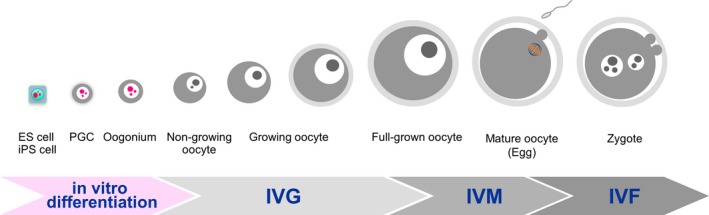
Figure 1
Full legend
Schematic flow of the in vitro differentiation of ES cells and iPS cells, and in vitro growth (IVG), in vitro maturation (IVM), and in vitro fertilization (IVF) of mammalian oocytes. ES cell: embryonic stem cell; iPS cell: induced pluripotent stem cell; and PGC: primordial germ cell
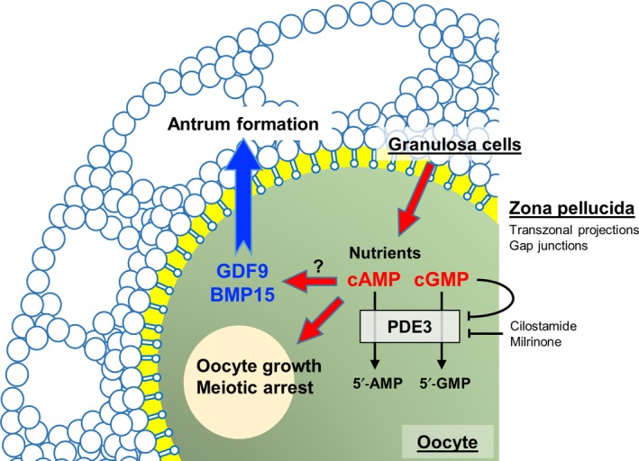
Figure 2
Full legend
Schematic model of communication between oocytes and granulosa cells. Nutrients, cAMP, and cGMP are transported from granulosa cells through their transzonal projections and gap junctions to oocytes to support the growth and meiotic arrest of oocytes. The oocyte‐derived growth factors GDF9 and BMP15 promote granulosa cell proliferation and differentiation, and antrum formation. Inhibition of oocyte PDE3 increases the expressions of GDF9 and BMP15, which in turn promote antrum formation. PDE3: phosphodiesterase 3; cAMP: cyclic adenosine 3′, 5′‐monophosphate; cGMP: cyclic guanosine 3′, 5′‐monophosphate; 5′‐AMP: 5′‐adenosine monophosphate; 5′‐GMP: 5′‐guanosine monophosphate; and PDE3 inhibitors: cilostamide and milrinone
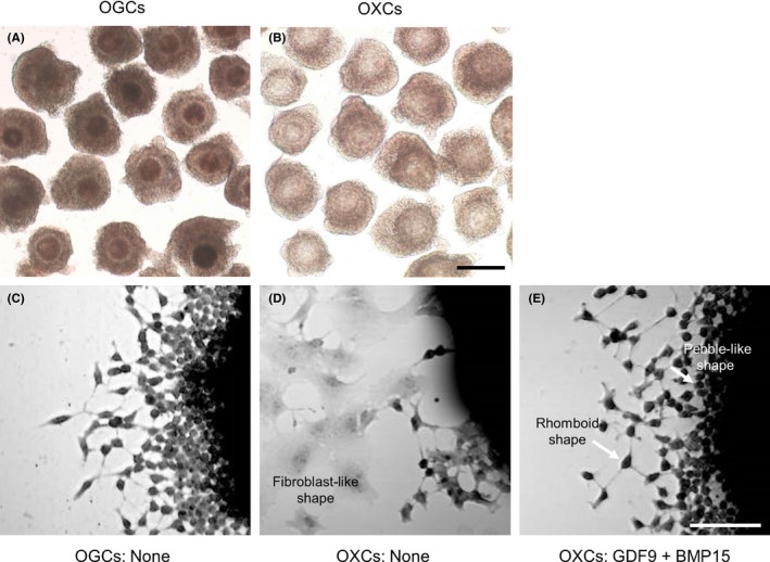
Figure 3
Full legend
Preparation and culture of bovine oocyte‐granulosa cell complexes (OGCs). OGCs containing growing oocytes (A) were collected from small antral follicles (1.2–1.8 mm in diameter). From OGCs, oocyte‐ectomized complexes (OXCs: B) were prepared by removing the oocyte cytoplasm with the germinal vesicle. After 5 days of culture, the complexes were fixed and stained with hematoxylin and eosin Y (C–E). In OXCs (D), outgrowing granulosa cells differentiated into fibroblast‐like cells. GDF9 and BMP15 reduced fibroblast‐like cells in OXCs, while rhomboid‐shaped and pebble‐like cells were observed (E), which were similar to those in control OGCs cultured without these factors (C). The scale bar in A and B represents 200 μm; that in C–E represents 50 μm
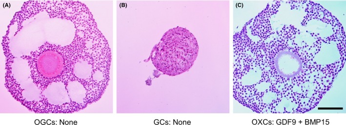
Figure 4
Full legend
Representative images of histological sections from cultured bovine growing oocyte‐granulosa cell complexes (OGCs). OGCs containing growing oocytes were collected form bovine small antral follicles as described in the footnote of Figure 3. From OGCs, oocyte‐ectomized complexes (OXCs) and granulosa cell complexes (GCs) without oocytes were prepared. After culture, the complexes were fixed, sectioned, and stained with hematoxylin and eosin Y. OGCs developed antrum‐like structures in vitro (A), whereas GCs (B) and OXCs (not shown) did not. However, OXCs (C) developed antrum‐like structures when they were cultured with GDF9 and BMP15. The scale bar represents 100 μm
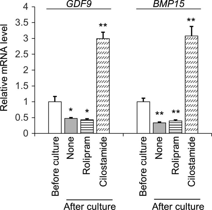
Figure 5
Full legend
Relative expression levels of GDF9 and BMP15 mRNAs in bovine growing oocytes after 5 days of culture with phosphodiesterase (PDE) inhibitors were assessed by qPCR. Oocytes cultured without a PDE inhibitor were marked as “None.” Rolipram (25 µM) was used as a PDE4 inhibitor and cilostamide (5 µM) as a PDE3 inhibitor. Bovine β‐ACTIN was used as internal control. Data are presented as fold changes relative to the control group (before culture) and are shown as the mean ± SEM from at least three replications. *P <.05; **P <.01
References133
- 1.
Byskov AG. Primordial germ cells and regulation of meiosis In: Austin CR, Short RV eds. Reproduction in mammals: 1. Germ cells and fertilization, 2nd edn Cambridge, UK: Cambridge University Press; 1982:1‐16.
Not in our library
- 2.
Mayo K, Jameson L, Woodruff TK. Eggs in the nest. Endocrinology. 2007;148:3577‐3579.
Not in our library10.1210/en.2007-0590 (opens in a new tab)
- 3.
Mhawi AJ, Kaňka J, Motlík J. Follicle and oocyte growth in early postnatal calves: cytochemical, autoradiographical and electron microscopical studies. Reprod Nutr Dev. 1991;31:115‐126.
Not in our library10.1051/rnd:19910201 (opens in a new tab)
- 4.
Wigglesworth K, Lee KB, Emori C, et al. Transcriptomic diversification of developing cumulus and mural granulosa cells in mouse ovarian follicles. Biol Reprod. 2015;92:1‐14.
Not in our library10.1095/biolreprod.114.121756 (opens in a new tab)
- 5.
Baird DT, Fraser IS. Disorders of the hypothalamic‐pituitary‐ovarian axis. Clin Endocrinol Metab. 1973;2:469‐488.
Not in our library10.1016/s0300-595x(73)80010-6 (opens in a new tab)
- 6.
Richards JS. Hormonal control of gene expression in the ovary. Endocr Rev. 1994;15:725‐751.
Not in our library10.1210/edrv-15-6-725 (opens in a new tab)
- 7.
Oktay K, Briggs D, Gosden RG. Ontogeny of follicle stimulating hormone receptor gene expression in isolated human ovarian follicles. J Clin Endocrinol. 1997;82:3748‐3751.
Not in our library10.1210/jcem.82.11.4346 (opens in a new tab)
- 8.
McGee E, Spears N, Minami S, et al. Preantral ovarian follicles in serum‐free culture: suppression of apoptosis after activation of the cyclic guanosine 3́,5́‐monophosphate pathway and stimulation of growth and differentiation by follicle‐stimulating hormone. Endocrinology. 1997;138:2417‐2424.
Not in our library10.1210/endo.138.6.5164 (opens in a new tab)
- 9.
Hsueh AJ, Billig H, Tsafriri A. Ovarian follicle atresia: a hormonally controlled apoptotic process. Endocr Rev. 1994;15:707‐724.
Not in our library10.1210/edrv-15-6-707 (opens in a new tab)
- 10.
Anderson E, Albertini DF. Gap junctions between the oocyte and companion follicle cells in the mammalian ovary. J Cell Biol. 1976;71:680‐686.
Not in our library10.1083/jcb.71.2.680 (opens in a new tab)
- 11.
Racowsky C, Satterlie RA. Metabolic, fluorescent dye and electrical coupling between hamster oocytes and cumulus cells during meiotic maturation in vivo and in vitro. Dev Biol. 1985;108:191‐202.
Not in our library10.1016/0012-1606(85)90022-3 (opens in a new tab)
- 12.
Eppig JJ. Intercommunication between mammalian oocytes and companion somatic cells. BioEssays. 1991;13:569‐574.
Not in our library10.1002/bies.950131105 (opens in a new tab)
- 13.
Eppig JJ. Mouse oocyte development in vitro with various culture systems. Dev Biol. 1977;60:371‐388.
Not in our library10.1016/0012-1606(77)90135-x (opens in a new tab)
- 14.
Eppig JJ, O’Brien MJ. Development in vitro of mouse oocytes from primordial follicles. Biol Reprod. 1996;54:197‐207.
Not in our library10.1095/biolreprod54.1.197 (opens in a new tab)
- 15.
Morohaku K, Tanimoto R, Sasaki K, et al. Complete in vitro generation of fertile oocytes from mouse primordial germ cells. Proc Natl Acad Sci USA. 2016;113:9021‐9026.
Not in our library10.1073/pnas.1603817113 (opens in a new tab)
- 16.
Hayashi K, Ohta H, Kurimoto K, Aramaki S, Saitou M. Reconstitution of the mouse germ cell specification pathway in culture by pluripotent stem cells. Cell. 2011;146:519‐532.
Not in our library10.1016/j.cell.2011.06.052 (opens in a new tab)
- 17.
Hayashi K, Ogushi S, Kurimoto K, Shimamoto S, Ohta H, Saitou M. Offspring from oocytes derived from in vitro primordial germ cell‐like cells in mice. Science. 2012;338:971‐975.
Not in our library10.1126/science.1226889 (opens in a new tab)
- 18.
Hikabe O, Hamazaki N, Nagamatsu GO, et al. Reconstitution in vitro of the entire cycle of the mouse female germ line. Nature. 2016;539:299‐303.
Not in our library10.1038/nature20104 (opens in a new tab)
- 19.
Hirao Y, Itoh T, Shimizu M, et al. In vitro growth and development of bovine oocyte‐granulosa cell complexes on the flat substratum: effects of high polyvinylpyrrolidone concentration in culture medium. Biol Reprod. 2004;70:83‐91.
Not in our library10.1095/biolreprod.103.021238 (opens in a new tab)
- 20.
Dong J, Albertini DF, Nishimori K, Kumar TR, Lu N, Matzuk MM. Growth differentiation factor‐9 is required during early ovarian folliculogenesis. Nature. 1996;383:531‐535.
Not in our library10.1038/383531a0 (opens in a new tab)
- 21.
Yan C, Wang P, DeMayo J, et al. Synergistic roles of bone morphogenetic protein 15 and growth differentiation factor 9 in ovarian function. Mol Endocrinol. 2001;15:854‐866.
Not in our library10.1210/mend.15.6.0662 (opens in a new tab)
- 22.
Matzuk MM, Burns KH, Viveiros MM, Eppig JJ. Intercellular communication in the mammalian ovary: oocytes carry the conversation. Science. 2002;296:2178‐2180.
Not in our library10.1126/science.1071965 (opens in a new tab)
- 23.
Hirao Y. Conditions affecting growth and developmental competence of mammalian oocytes in vitro. Anim Sci J. 2011;82:187‐197.
Not in our library10.1111/j.1740-0929.2010.00870.x (opens in a new tab)
- 24.
Hirao Y. Isolation of ovarian components essential for growth and development of mammalian oocytes in vitro. J Reprod Dev. 2012;58:167‐174.
Not in our library10.1262/jrd.2011-052 (opens in a new tab)
- 25.
Hirao Y. Oocyte growth in vitro: potential model for studies of oocyte‐granulosa cell interactions. Reprod Med Biol. 2011;11:1‐9.
Not in our library10.1007/s12522-011-0096-3 (opens in a new tab)
- 26.
Brower PT, Schultz RM. Intercellular communication between granulosa cells and mouse oocytes: existence and possible nutritional role during oocyte growth. Dev Biol. 1982;90:144‐153.
Not in our library10.1016/0012-1606(82)90219-6 (opens in a new tab)
- 27.
Baena V, Terasaki M. Three‐dimensional organization of transzonal projections and other cytoplasmic extensions in the mouse ovarian follicle. Sci Rep. 2019;9:1262.
Not in our library10.1038/s41598-018-37766-2 (opens in a new tab)
- 28.
Macaulay AD, Gilbert I, Caballero J, et al. The gametic synapse: RNA transfer to the bovine oocyte. Biol Reprod. 2014;91:1‐12.
Not in our library10.1095/biolreprod.114.119867 (opens in a new tab)
- 29.
Albertini DF, Rider DF. Pattern of intercellular connectivity in the mammalian cumulus‐oocyte complex. Microsc Res Tech. 1994;27:125‐133.
Not in our library10.1002/jemt.1070270206 (opens in a new tab)
- 30.
El‐Hayek S, Yang Q, Abbassi L, FitzHarris G, Clarke HJ. Mammalian oocytes locally remodel follicular architecture to provide the foundation for germline‐soma communication. Curr Biol. 2018;28:1124‐1131.
Not in our library10.1016/j.cub.2018.02.039 (opens in a new tab)
- 31.
Eppig JJ. Reproduction: oocytes call, granulosa cells connect. Curr Biol. 2018;28:354‐356.
Not in our library10.1016/j.cub.2018.03.005 (opens in a new tab)
- 32.
Motta PM, Makabe S, Naguro T, Correr S. Oocyte follicle cells association during development of human ovarian follicle. A study by high resolution scanning and transmission electron microscopy. Arch Histol Cytol. 1994;57:369‐394.
Not in our library10.1679/aohc.57.369 (opens in a new tab)
- 33.
Makita M, Miyano T. Steroid hormones promote bovine oocyte growth and connection with granulosa cells. Theriogenology. 2014;82:605‐612.
Not in our library10.1016/j.theriogenology.2014.05.020 (opens in a new tab)
- 34.
Cayo‐Colca IS, Yamagami Y, Phan TC, Miyano T. A combination of FSH and dibutyryl cyclic AMP promote growth and acquisition of meiotic competence of oocytes from early porcine antral follicles. Theriogenology. 2011;75:1602‐1612.
Not in our library10.1016/j.theriogenology.2010.12.023 (opens in a new tab)
- 35.
Simon AM, Goodenough DA. Diverse functions of vertebrate gap junctions. Trends Cell Biol. 1998;8:477‐483.
Not in our library10.1016/s0962-8924(98)01372-5 (opens in a new tab)
- 36.
Willecke K, Eiberger J, Degen J, et al. Structural and functional diversity of connexin genes in the mouse and human genome. Biol Chem. 2002;383:725‐737.
Not in our library10.1515/bc.2002.076 (opens in a new tab)
- 37.
Kidder GM, Vanderhyden BC. Bidirectional communication between oocytes and follicle cells: ensuring oocyte development competence. Can J Physiol Pharmacol. 2010;88:399‐413.
Not in our library10.1139/y10-009 (opens in a new tab)
- 38.
Kidder GM, Mhawi AA. Gap junctions and ovarian folliculogenesis. Reproduction. 2002;123:613‐620.
Not in our library10.1530/rep.0.1230613 (opens in a new tab)
- 39.
Teilmann SC. Differential expression and localisation of connexin‐37 and connexin‐43 in follicles of different stages in the 4‐week‐old mouse ovary. Mol Cell Endocrinol. 2005;234:27‐35.
Not in our library10.1016/j.mce.2004.10.014 (opens in a new tab)
- 40.
Johnson ML, Redmer DA, Reynolds LP, Grazul‐Bilska AT. Expression of gap junctional proteins connexin 43, 32, and 26 throughout follicular development and atresia in cows. Endocrine. 1999;10:43‐51.
Not in our library10.1385/endo:10:1:43 (opens in a new tab)
- 41.
Johnson ML, Redmer DA, Reynolds LP, Bilski JJ, Grazul‐Bilska AT. Gap junctional intercellular communication of bovine granulosa and thecal cells from antral follicles: effects of luteinizing hormone and follicle stimulating hormone. Endocrine. 2002;18:261‐270.
Not in our library10.1385/endo:18:3:261 (opens in a new tab)
- 42.
Nuttinck F, Peynot N, Humblot P, Massip A, Dessy F, Fléchon JE. Comparative immunohistochemical distribution of connexin 37 and connexin 43 throughout folliculogenesis in the bovine ovary. Mol Reprod Dev. 2000;57:60‐66.
Not in our library10.1002/1098-2795(200009)57:1<60::aid-mrd9>3.0.co;2-6 (opens in a new tab)
- 43.
Wassarman PM. Channels of communication in the ovary. Nat Cell Biol. 2002;4:7‐9.
Not in our library10.1038/ncb-nm-fertilitys7 (opens in a new tab)
- 44.
Simon AM, Goodenough DA, Li E, Paul DL. Female infertility in mice lacking connexin37. Nature. 1997;385:525‐529.
Not in our library10.1038/385525a0 (opens in a new tab)
- 45.
Carabatsos MJ, Sellitto C, Goodenough DA, Albertini DF. Oocyte‐granulosa cell heterologous gap junctions are required for the coordination of nuclear and cytoplasmic meiotic competence. Dev Biol. 2000;226:167‐179.
Not in our library10.1006/dbio.2000.9863 (opens in a new tab)
- 46.
Colonna R, Mangia F. Mechanisms of amino acid uptake in cumulus‐enclosed mouse oocytes. Biol Reprod. 1983;28:797‐803.
Not in our library10.1095/biolreprod28.4.797 (opens in a new tab)
- 47.
Haghighat N, Van Winkle LJ. Developmental change in follicular cell‐enhanced amino acid uptake into mouse oocytes that depends on intact gap junctions and transport system Gly. J Exp Zool. 1990;253:71‐82.
Not in our library10.1002/jez.1402530110 (opens in a new tab)
- 48.
Donahue RP, Stern S. Follicular cell support of oocyte maturation: production of pyruvate in vitro. J Reprod Fertil. 1968;17:395‐398.
Not in our library10.1530/jrf.0.0170395 (opens in a new tab)
- 49.
Pelland AM, Corbett HE, Baltz JM. Amino acid transport mechanisms in mouse oocytes during growth and meiotic maturation. Biol Reprod. 2009;81:1041‐1054.
Not in our library10.1095/biolreprod.109.079046 (opens in a new tab)
- 50.
Purcell SH, Moley KH. Glucose transporters in gametes and preimplantation embryos. Trends Endrocrinol Metab. 2009;20:483‐489.
Not in our library10.1016/j.tem.2009.06.006 (opens in a new tab)
- 51.
Augustin R, Pocar P, Navarrete‐Santos A, et al. Glucose transporter expression is developmentally regulated in in vitro derived bovine preimplantation embryos. Mol Reprod Dev. 2001;60:370‐376.
Not in our library10.1002/mrd.1099 (opens in a new tab)
- 52.
Pisani LF, Antonini S, Pocar P, et al. Effects of pre‐mating nutrition on mRNA levels of developmentally relevant genes in sheep oocytes and granulosa cells. Reproduction. 2008;136:303‐312.
Not in our library10.1530/rep-07-0394 (opens in a new tab)
- 53.
Dan‐Goor M, Sasson S, Davarashvili A, Almagor M. Expression of glucose transporter and glucose uptake in human oocytes and preimplantation embryos. Hum Reprod. 1997;12:2508‐2510.
Not in our library10.1093/humrep/12.11.2508 (opens in a new tab)
- 54.
Zheng P, Vassena R, Latham KE. Effects of in vitro oocyte maturation and embryo culture on the expression of glucose transporters, glucose metabolism and insulin signaling genes in rhesus monkey oocytes and preimplantation embryos. Mol Hum Reprod. 2007;13:361‐371.
Not in our library10.1093/molehr/gam014 (opens in a new tab)
- 55.
Brinster RL. Oxidation of pyruvate and glucose by oocytes of the mouse and rhesus monkey. J Reprod Fertil. 1971;24:187‐191.
Not in our library10.1530/jrf.0.0240187 (opens in a new tab)
- 56.
Steeves TE, Gardner DK. Metabolism of glucose, pyruvate and glutamine during the maturation of oocytes derived from pre‐pubertal and adult cows. Mol Reprod Dev. 1999;54:92‐101.
Not in our library10.1002/(sici)1098-2795(199909)54:1<92::aid-mrd14>3.0.co;2-a (opens in a new tab)
- 57.
Sutton‐McDowall ML, Gilchrist RB, Thompson JG. The pivotal role of glucose metabolism in determining oocyte developmental competence. Reproduction. 2010;139:685‐695.
Not in our library10.1530/rep-09-0345 (opens in a new tab)
- 58.
Cetica P, Pintos L, Dalvit G, Beconi M. Activity of key enzymes involved in glucose and triglyceride catabolism during bovine oocyte maturation in vitro. Reproduction. 2002;124:675‐681.
Not in our library
- 59.
Leese HJ, Barton AM. Production of pyruvate by isolated mouse cumulus cells. J Exp Zool. 1985;234:231‐236.
Not in our library10.1002/jez.1402340208 (opens in a new tab)
- 60.
Biggers JD, Whittingham DG, Donahue RP. The pattern of energy metabolism in the mouse oocyte and zygote. Proc Natl Acad Sci USA. 1967;58:560‐567.
Not in our library10.1073/pnas.58.2.560 (opens in a new tab)
- 61.
Honda A, Hirose M, Inoue K, et al. Large‐scale production of growing oocytes in vitro from neonatal mouse ovaries. Int J Dev Biol. 2009;53:605‐613.
Not in our library10.1387/ijdb.082607ah (opens in a new tab)
- 62.
Fair T, Hyttel P, Greve T. Bovine oocyte diameter in relation to maturational competence and transcriptional activity. Mol Reprod Dev. 1995;42:437‐442.
Not in our library10.1002/mrd.1080420410 (opens in a new tab)
- 63.
Sorensen RA, Wassarman PM. Relationship between growth and meiotic maturation of the mouse oocyte. Dev Biol. 1976;50:531‐536.
Not in our library10.1016/0012-1606(76)90172-x (opens in a new tab)
- 64.
Pincus G, Enzmann EV. The comparative behavior of mammalian eggs in vivo and in vitro: I. the activation of ovarian eggs. J Exp Med. 1935;62:665‐675.
Not in our library10.1084/jem.62.5.665 (opens in a new tab)
- 65.
Leibfried L, First NL. Follicular control of meiosis in the porcine oocyte. Biol Reprod. 1980;23:705‐709.
Not in our library10.1095/biolreprod23.4.705 (opens in a new tab)
- 66.
Cho WK, Stern S, Biggers JD. Inhibitory effect of dibutyryl cAMP on mouse oocyte maturation in vitro. Exp Zool. 1974;187:383‐386.
Not in our library10.1002/jez.1401870307 (opens in a new tab)
- 67.
Dekel N, Beers WH. Rat oocyte maturation in vitro: relief of cyclic AMP inhibition by gonadotropins. Proc Natl Acad Sci USA. 1978;75:4369‐4373.
Not in our library10.1073/pnas.75.9.4369 (opens in a new tab)
- 68.
Dekel N, Beers WH. Development of the rat oocyte in vitro: inhibition and induction of maturation in the presence or absence of the cumulus oophorus. Dev Biol. 1980;75:247‐254.
Not in our library10.1016/0012-1606(80)90160-8 (opens in a new tab)
- 69.
Norris RP, Ratzan WJ, Freudzon M, et al. Cyclic GMP from the surrounding somatic cells regulates cyclic AMP and meiosis in the mouse oocyte. Development. 2009;136:1869‐1878.
Not in our library10.1242/dev.035238 (opens in a new tab)
- 70.
Schultz RM, Montgomery RR, Ward‐Bailey PF, Eppig JJ. Regulation of oocyte maturation in the mouse: possible roles of intracellular communication, cAMP and testosterone. Dev Biol. 1983;95:294‐304.
Not in our library10.1016/0012-1606(83)90030-1 (opens in a new tab)
- 71.
Sirard MA, First NL In vitro inhibition of oocyte nuclear maturation in the bovine. Biol Reprod. 1988;39:229‐234.
Not in our library10.1095/biolreprod39.2.229 (opens in a new tab)
- 72.
Tsafriri A, Chun S‐Y, Zhang R, Hsueh A, Conti M. Oocyte maturation involves compartmentalization and opposing changes of cAMP levels in follicular somatic and germ cells: studies using selective phosphodiesterase inhibitors. Dev Biol. 1996;178:393‐402.
Not in our library10.1006/dbio.1996.0226 (opens in a new tab)
- 73.
Shitsukawa K, Andersen C, Richard F, et al. Cloning and characterization of the cyclic guanosine monophosphate‐inhibited phosphodiesterase PDE3A expressed in mouse oocyte. Biol Reprod. 2001;65:188‐196.
Not in our library10.1095/biolreprod65.1.188 (opens in a new tab)
- 74.
Sasseville M, Côte N, Guillemette C, Richard F. New insight into the role of phosphodiesterase 3A in porcine oocyte maturation. BMC Dev Biol. 2006;6:47.
Not in our library10.1186/1471-213x-6-47 (opens in a new tab)
- 75.
Thomas RE, Armstrong DT, Gilchrist RB. Differential effects of specific phosphodiesterase isoenzyme inhibitors on bovine oocyte meiotic maturation. Dev Biol. 2002;244:215‐225.
Not in our library10.1006/dbio.2002.0609 (opens in a new tab)
- 76.
Robinson JW, Zhang M, Shuhaibar LC, et al. Luteinizing hormone reduces the activity of the NPR2 guanylyl cyclase in mouse ovarian follicles, contributing to the cyclic GMP decrease that promotes resumption of meiosis in oocytes. Dev Biol. 2012;366:308‐316.
Not in our library10.1016/j.ydbio.2012.04.019 (opens in a new tab)
- 77.
Zhang M, Su Y‐Q, Sugiura K, Xia G, Eppig JJ. Granulosa cell ligand NPPC and its receptor NPR2 maintain meiotic arrest in mouse oocytes. Science. 2010;330:366‐369.
Not in our library10.1126/science.1193573 (opens in a new tab)
- 78.
Wigglesworth K, Lee K‐B, O'Brien MJ, Peng J, Matzuk MM, Eppig JJ. Bidirectional communication between oocytes and ovarian follicular somatic cells is required for meiotic arrest of mammalian oocytes. Proc Natl Acad Sci USA. 2013;110:3723‐3729.
Not in our library10.1073/pnas.1314829110 (opens in a new tab)
- 79.
Downs SM, Eppig JJ. Cyclic adenosine monophosphate and ovarian follicular fluid act synergistically to inhibit mouse oocyte maturation. Endocrinology. 1984;114:418‐427.
Not in our library10.1210/endo-114-2-418 (opens in a new tab)
- 80.
Downs SM, Coleman DL, Ward‐Bailey PF, Eppig JJ. Hypoxanthine is the principal inhibitor of murine oocyte maturation in a low molecular weight fraction of porcine follicular fluid. Proc Natl Acad Sci USA. 1985;82:454‐458.
Not in our library10.1073/pnas.82.2.454 (opens in a new tab)
- 81.
Eppig JJ, Ward‐Bailey PF, Coleman DL. Hypoxanthine and adenosine in murine ovarian follicular fluid: concentrations and activity in maintaining oocyte meiotic arrest. Biol Reprod. 1985;33:1041‐1049.
Not in our library10.1095/biolreprod33.5.1041 (opens in a new tab)
- 82.
Miyano T, Ebihara M, Goto Y, Hirao Y, Nagai T, Kato S. Inhibitory action of hypoxanthine on meiotic resumption of denuded pig follicular oocytes in vitro. J Exp Zool. 1995;273:70‐75.
Not in our library10.1002/jez.1402730109 (opens in a new tab)
- 83.
Moritake S, Hirao Y, Miyano T. Hypoxanthine promotes the acquisition of meiotic competence in pig oocytes from early antral follicles during growth culture. J Mamm Ova Res. 2002;19:39‐45.
Not in our library
- 84.
Harada M, Miyano T, Matsumura K, Osaki S, Miyake M, Kato S. Bovine oocytes from early antral follicles grow to meiotic competence in vitro: effect of FSH and hypoxanthine. Theriogenology. 1996;48:743‐755.
Not in our library10.1016/s0093-691x(97)00298-7 (opens in a new tab)
- 85.
Rao MC, Midgley AR Jr, Richards JS. Hormonal regulation of ovarian cellular proliferation. Cell. 1978;14:71‐78.
Not in our library10.1016/0092-8674(78)90302-1 (opens in a new tab)
- 86.
Herath CB, Yamashita M, Watanabe G, et al. Regulation of follicle‐stimulating hormone secretion by estradiol and dimeric inhibins in the infantile rat. Biol Reprod. 2001;65:1623‐1633.
Not in our library10.1095/biolreprod65.6.1623 (opens in a new tab)
- 87.
Drummond AE. TGFβ signalling in the development of ovarian function. Cell Tissue Res. 2005;322:107‐115.
Not in our library10.1007/s00441-005-1153-1 (opens in a new tab)
- 88.
Orr‐Urtreger A, Avivi A, Zimmer Y, et al. Developmental expression of c‐kit, a proto‐oncogene encoded by the W locus. Development. 1990;109:911‐923.
Not in our library10.1242/dev.109.4.911 (opens in a new tab)
- 89.
Horie K, Fujita J, Takakura K, et al. The expression of c‐kit protein in human adult and fetal tissues. Human Reprod. 1993;8:1955‐1962.
Not in our library10.1093/oxfordjournals.humrep.a137967 (opens in a new tab)
- 90.
Ismail R, Okawara J, Fryer JN, Vanderhyden BC. Hormonal regulation of the ligand for c‐kit in the rat ovary and its effects on spontaneous oocyte meiotic maturation. Mol Reprod Dev. 1996;43:458‐469.
Not in our library10.1002/(sici)1098-2795(199604)43:4<458::aid-mrd8>3.0.co;2-o (opens in a new tab)
- 91.
Manova K, Huang EJ, Angeles M, et al. The expression pattern of the c‐kit ligand in gonads of mice supports a role for the c‐kit receptor in oocyte growth and in proliferation of spermatogonia. Dev Biol. 1993;157:85‐99.
Not in our library10.1006/dbio.1993.1114 (opens in a new tab)
- 92.
Laitinen M, Rutanen EM, Ritvos O. Expression of c‐kit ligand messenger ribonucleic acids in human ovaries and regulation of their steady state levels by gonadotropins in cultured granulosa‐luteal cells. Endocrinology. 1995;136:4407‐4414.
Not in our library10.1210/endo.136.10.7545103 (opens in a new tab)
- 93.
Packer AI, Hsu YC, Besmer P, Bachvarova RF. The ligand of the c‐kit receptor promotes oocyte growth. Dev Biol. 1994;161:194‐205.
Not in our library10.1006/dbio.1994.1020 (opens in a new tab)
- 94.
Parrott JA, Skinner MK. Kit‐ligand/stem cell factor induces primordial follicle development and initiates folliculogenesis. Endocrinology. 1999;140:4262‐4271.
Not in our library10.1210/endo.140.9.6994 (opens in a new tab)
- 95.
Moniruzzaman M, Sakamaki K, Akazawa Y, Miyano T. Oocyte growth and follicular development in KIT‐deficient Fas‐knockout mice. Reproduction. 2007;133:117‐125.
Not in our library10.1530/rep-06-0161 (opens in a new tab)
- 96.
John GB, Shidler MJ, Besmer P, Castrillon DH. Kit signaling via PI3K promotes ovarian follicle maturation but is dispensable for primordial follicle activation. Dev Biol. 2009;331:292‐299.
Not in our library10.1016/j.ydbio.2009.05.546 (opens in a new tab)
- 97.
Saatcioglu HD, Cuevas I, Castrillon DH. Control of oocyte reawakening by kit. PLoS Genet. 2016;12:e1006215.
Not in our library10.1371/journal.pgen.1006215 (opens in a new tab)
- 98.
El‐fouly MA, Cook B, Nekola M, Nalbandov AV. Nalbandov AV. Role of the ovum in follicular luteinization. Endocrinology. 1970;87:286‐293.
Not in our library
- 99.
Nekola MV, Nalbandov AV. Morphological changes of rat follicular cells as influenced by oocytes. Biol Reprod. 1971;4:154‐160.
Not in our library10.1093/biolreprod/4.2.154 (opens in a new tab)
- 100.
Vanderhyden BC, Caron PJ, Buccione R, Eppig JJ. Developmental pattern of the secretion of cumulus expansion‐enabling factor by mouse oocytes and the role of oocytes in promoting granulosa cell differentiation. Dev Biol. 1990;140:307‐317.
Not in our library10.1016/0012-1606(90)90081-s (opens in a new tab)
- 101.
Buccione R, Vanderhyden BC, Caron PJ, Eppig JJ. FSH‐induced expansion of the mouse cumulus oophorus in vitro is dependent upon a specific factor(s) secreted by the oocyte. Dev Biol. 1990;138:16‐25.
Not in our library10.1016/0012-1606(90)90172-f (opens in a new tab)
- 102.
Ralph JH, Telfer EE, Wilmut I. Bovine cumulus cell expansion does not depend on the presence of an oocyte secreted factor. Mol Reprod Dev. 1995;42:248‐253.
Not in our library10.1002/mrd.1080420214 (opens in a new tab)
- 103.
Prochazka R, Nagyova E, Rimkevicova Z, Nagai T, Kikuchi K, Motlik J. Lack of effect of oocytectomy on expansion of the porcine cumulus. J Reprod Fert. 1991;93:569‐576.
Not in our library10.1530/jrf.0.0930569 (opens in a new tab)
- 104.
Shen X, Miyano T, Kato S. Promotion of follicular antrum formation by pig oocytes in vitro. Zygote. 1998;6:47‐54.
Not in our library10.1017/s0967199400005074 (opens in a new tab)
- 105.
Sugiura K, Pendola FL, Eppig JJ. Oocyte control of metabolic cooperativity between oocytes and companion granulosa cells: energy metabolism. Dev Biol. 2005;279:20‐30.
Not in our library10.1016/j.ydbio.2004.11.027 (opens in a new tab)
- 106.
Hanrahan JP, Gregan SM, Mulsant P, et al. Mutations in the genes for oocyte‐derived growth factors GDF9 and BMP15 are associated with both increased ovulation rate and sterility in Cambridge and Belclare sheep (Ovis aries). Biol Reprod. 2004;70:900‐909.
Not in our library10.1095/biolreprod.103.023093 (opens in a new tab)
- 107.
Juengel JL, Hudson NL, Berg M, et al. Effects of active immunization against growth differentiation factor 9 and/or bone morphogenetic protein 15 on ovarian function in cattle. Reproduction. 2009;138:107‐114.
Not in our library10.1530/rep-09-0009 (opens in a new tab)
- 108.
Elvin JA, Clark AT, Wang P, Wolfman NM, Matzuk MM. Paracrine actions of growth differentiation factor‐9 in the mammalian ovary. Mol Endocrinol. 1999;13:1035‐1048.
Not in our library10.1210/mend.13.6.0310 (opens in a new tab)
- 109.
Sugiura K, Su Y‐Q, Diaz FJ, et al. Oocyte‐derived BMP15 and FGFs cooperate to promote glycolysis in cumulus cells. Development. 2007;134:2593‐2603.
Not in our library10.1242/dev.006882 (opens in a new tab)
- 110.
McGrath SA, Esquela AF, Lee SJ. Oocyte‐specific expression of growth/differentiation factor‐9. Mol Endocrinol. 1995;9:131‐136.
Not in our library10.1210/mend.9.1.7760846 (opens in a new tab)
- 111.
Dube JL, Wang P, Elvin J, Lyons KM, Celeste AJ, Matzuk MM. The bone morphogenetic protein 15 gene is X‐linked and expressed in oocytes. Mol Endocrinol. 1998;12:1809‐1817.
Not in our library10.1210/mend.12.12.0206 (opens in a new tab)
- 112.
Hayashi M, McGee EA, Min G, et al. Recombinant growth differentiation factor‐9 (GDF‐9) enhances growth and differentiation of cultured early ovarian follicles. Endocrinology. 1999;140:1236‐1244.
Not in our library10.1210/endo.140.3.6548 (opens in a new tab)
- 113.
Jaatinen R, Laitinen MP, Vuojolainen K, et al. Localization of growth differentiation factor‐9 (GDF‐9) mRNA and protein in rat ovaries and cDNA cloning of rat GDF‐9 and its novel homolog GDF‐9B. Mol Cell Endocrinol. 1999;156:189‐193.
Not in our library10.1016/s0303-7207(99)00100-8 (opens in a new tab)
- 114.
Erickson GF, Shimasaki S. The spatiotemporal expression pattern of the bone morphogenetic protein family in rat ovary cell types during the estrous cycle. Reprod Biol Endocrinol. 2003;1:9.
Not in our library10.1186/1477-7827-1-9 (opens in a new tab)
- 115.
Bodensteiner KJ, Clay CM, Moeller CL, Sawyer HR. Molecular cloning of the ovine growth/differentiation factor‐9 gene and expression of growth/differentiation factor‐9 in ovine and bovine ovaries. Biol Reprod. 1999;60:381‐386.
Not in our library10.1095/biolreprod60.2.381 (opens in a new tab)
- 116.
Galloway SM, McNatty KP, Cambridge LM, et al. Mutations in an oocyte‐derived growth factor gene (BMP15) cause increased ovulation rate and infertility in a dosage‐sensitive manner. Nat Genet. 2000;25:279‐283.
Not in our library10.1038/77033 (opens in a new tab)
- 117.
Aaltonen J, Laitinen MP, Vuojolainen K, et al. Human growth differentiation factor 9 (GDF‐9) and its novel homolog GDF‐9B are expressed in oocytes during early folliculogenesis. J Clin Endocrinol Metab. 1999;84:2744‐2750.
Not in our library10.1210/jcem.84.8.5921 (opens in a new tab)
- 118.
Mottershead DG, Ritter LJ, Gilchrist RB. Signalling pathways mediating specific synergistic interactions between GDF9 and BMP15. Mol Hum Reprod. 2012;18:121‐128.
Not in our library10.1093/molehr/gar056 (opens in a new tab)
- 119.
Mottershead DG, Sugimura S, Al‐Musawi SL, et al. Cumulin, an oocyte‐secreted heterodimer of the transforming growth factor‐β family, is a potent activator of granulosa cells and improves oocyte quality. J Biol Chem. 2015;290:24007‐24020.
Not in our library10.1074/jbc.m115.671487 (opens in a new tab)
- 120.
Robker RL, Richards JS. Hormone‐induced proliferation and differentiation of granulosa cells: a coordinated balance of the cell cycle regulators cyclin D2 and p27Kip1. Mol Endocrinol. 1998;12:924‐940.
Not in our library10.1210/mend.12.7.0138 (opens in a new tab)
- 121.
Vanderhyden BC, Telfer EE, Eppig JJ. Mouse oocytes promote proliferation of granulosa cells from preantral and antral follicles in vitro. Biol Reprod. 1992;46:1196‐1204.
Not in our library10.1095/biolreprod46.6.1196 (opens in a new tab)
- 122.
McNatty KP, Juengel JL, Reader KL, et al. Bone morphogenetic protein 15 and growth differentiation factor 9 co‐operate to regulate granulosa cell function. Reproduction. 2005;129:473‐480.
Not in our library10.1530/rep.1.0511 (opens in a new tab)
- 123.
McIntosh CJ, Lun S, Lawrence S, et al. The proregion of mouse BMP15 regulates the cooperative interactions of BMP15 and GDF9. Biol Reprod. 2008;79:889‐896.
Not in our library10.1095/biolreprod.108.068163 (opens in a new tab)
- 124.
McNatty KP, Juengel JL, Reader KL, et al. Bone morphogenetic protein 15 and growth differentiation factor 9 co‐operate to regulate granulosa cell function in ruminants. Reproduction. 2005;129:481‐487.
Not in our library10.1530/rep.1.00517 (opens in a new tab)
- 125.
Knecht M, Amsterdam A, Catt K. The regulatory role of cyclic AMP in hormone‐induced of granulosa cell differentiation. J Biol Chem. 1981;256:10628‐10633.
Not in our library
- 126.
Lawrence TS, Ginzberg RD, Gilula NB, Beers WH. Hormonally induced cell shape changes in cultured rat ovarian granulosa cells. J Cell Biol. 1979;80:21‐36.
Not in our library10.1083/jcb.80.1.21 (opens in a new tab)
- 127.
Alam MH, Lee J, Miyano T. GDF9 and BMP15 induce development of antrum‐like structures by bovine granulosa cells without oocytes. J Reprod Dev. 2018;64:423‐431.
Not in our library10.1262/jrd.2018-078 (opens in a new tab)
- 128.
Kossowska‐Tomaszczuk K, De Geyter C, De Geyter M, et al. The multipotency of luteinizing granulosa cells collected from mature follicles. Stem Cells. 2009;27:210‐219.
Not in our library10.1634/stemcells.2008-0233 (opens in a new tab)
- 129.
Oki Y, Ono H, Motohashi T, Sugiura N, Nobusue H, Kano K. Dedifferentiated follicular granulosa cells derived from pig ovary can transdifferentiate into osteoblasts. Biochem J. 2012;447:239‐248.
Not in our library10.1042/bj20120172 (opens in a new tab)
- 130.
Nayudu PL, Osborn SM. Factors influencing the rate of preantral and antral growth of mouse ovarian follicles in vitro. J Reprod Fertil. 1992;95:349‐362.
Not in our library10.1530/jrf.0.0950349 (opens in a new tab)
- 131.
Hirao Y, Nagai T, Kubo M, Miyano T, Miyake M, Kato S In vitro growth and maturation of pig oocytes. J Reprod Fertil. 1994;100:333‐339.
Not in our library10.1530/jrf.0.1000333 (opens in a new tab)
- 132.
Gore‐Langton RE, Daniel SA. Follicle‐stimulating hormone and estradiol regulate antrum‐like reorganization of granulosa cells in rat preantral follicle cultures. Biol Reprod. 1990;43:65‐72.
Not in our library10.1095/biolreprod43.1.65 (opens in a new tab)
- 133.
Alam MH, Lee J, Miyano T. Inhibition of PDE3A sustains meiotic arrest and gap junction of bovine growing oocytes in in vitro growth culture. Theriogenology. 2018;118:110‐118.
Not in our library10.1016/j.theriogenology.2018.05.028 (opens in a new tab)
Reads
Read by people on this page
None yet.
Checked or read by AI through naulon
None yet.
A person counts once a day per paper. We keep no cookie and no address: a reader is a hash that changes every day. Any month under five reads shows no number.
Are you an author?
Is this your paper?
Claim it to add your note and be paid when agents cite it. Free.