Real‐time cardiovascular MR with spatio‐temporal artifact suppression using deep learning–proof of concept in congenital heart disease
Andreas Hauptmann, Simon Arridge, Felix Lucka and 2 more
Magnetic Resonance in Medicine · 2018 · CC BY
doi:10.1002/mrm.27480 (opens in a new tab)
We email you when a new notice or a question about this paper appears.
Abstract
Purpose. Real‐time assessment of ventricular volumes requires high acceleration factors. Residual convolutional neural networks (CNN) have shown potential for removing artifacts caused by data undersampling. In this study, we investigated the ability of CNNs to reconstruct highly accelerated radial real‐time data in patients with congenital heart disease (CHD).
Methods. A 3D (2D plus time) CNN architecture was developed and trained using synthetic training data created from previously acquired breath hold cine images from 250 CHD patients. The trained CNN was then used to reconstruct actual real‐time, tiny golden angle (tGA) radial SSFP data (13 × undersampled) acquired in 10 new patients with CHD. The same real‐time data was also reconstructed with compressed sensing (CS) to compare image quality and reconstruction time. Ventricular volume measurements made using both the CNN and CS reconstructed images were compared to reference standard breath hold data.
Results. It was feasible to train a CNN to remove artifact from highly undersampled radial real‐time data. The overall reconstruction time with the CNN (including creation of aliased images) was shown to be >5 × faster than the CS reconstruction. In addition, the image quality and accuracy of biventricular volumes measured from the CNN reconstructed images were superior to the CS reconstructions.
Conclusion. This article has demonstrated the potential for the use of a CNN for reconstruction of real‐time radial data within the clinical setting. Clinical measures of ventricular volumes using real‐time data with CNN reconstruction are not statistically significantly different from gold‐standard, cardiac‐gated, breath‐hold techniques.
Abstract licence: CC BY
No text here
Its licence does not allow us to show the full text. Read it at PubMed Central:
Read at PubMed Central (opens in a new tab)Figures7
From PubMed Central, U.S. National Library of Medicine. Licence: CC BY.
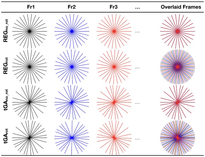
Figure 1
Full legend
The 4 radial sampling patterns tested in this study. Fully sampled k‐space requires 182 uniformly spaced radial spokes. An acceleration factor of 13 was used, resulting in 14 radial spokes acquired per frame, regardless of sampling pattern. REGno_rot, regular angle scheme (equal angular spacing of ~12.9o between each profile) with no rotation between frames. REGrot, regular angle scheme with regular rotation (of ~0.99o) between frames so that 13 consecutive frames constitutes a fully sampled k‐space. tGAno_rot, tiny golden angle scheme with no rotation between frames. tGArot, continuously rotating tiny golden angle sampling
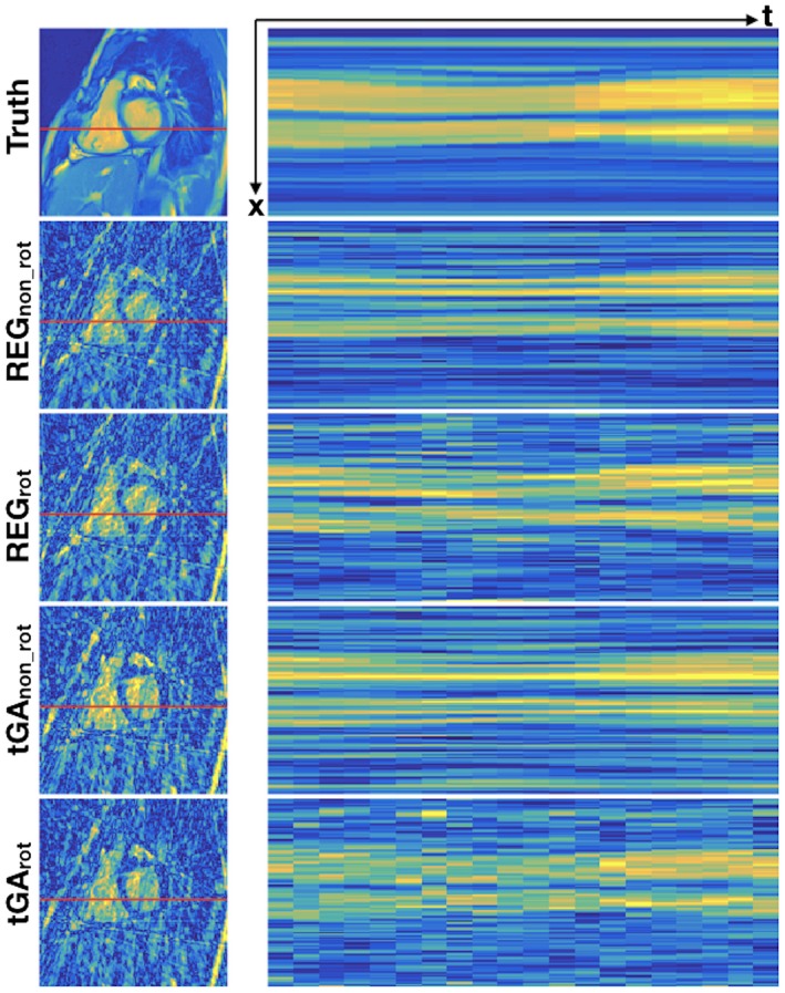
Figure 2
Full legend
Typical artifact produced from the 4 radial sampling patterns tested in this study. Left: a single frame from the cine. Right: 1 line through the ventricles (shown in red) through time. REGno_rot produces regular aliases with no variation through time. REGrot produces regular aliases in space that change position over time. tGAno_rot produces spatially incoherent aliases that do not vary through time. tGArot produces spatially incoherent aliases that vary through time
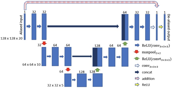
Figure 3
Full legend
The chosen residual U‐Net structure for spatio‐temporal deep de‐aliasing. The input is given by the aliased reconstruction from undersampled data. The numbers on top of the blue bars denote the number of channels for each layer. The resolution for each multilevel decomposition is shown in gray on the left. Each convolutional layer is equipped with a rectified linear unit as nonlinearity, given by ReLU(X) = max(x,0)
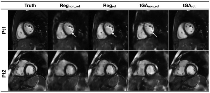
Figure 4
Full legend
Examples images from 2 “synthetic real‐time” test data sets reconstructed from the 4 different sampling patterns, using correspondingly trained residual U‐Net's. Pt1 shows total loss of some papillary muscles (indicated with arrow) compared to the truth data, with all trajectories except tGArot. Pt2 shows blurring across the ventricle, particularly seen with the non‐rotating trajectories (see Supporting Information Video S2 for corresponding movie)
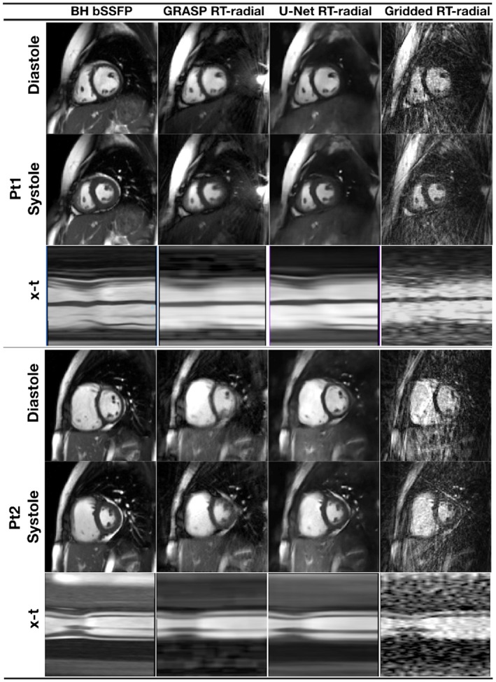
Figure 5
Full legend
Example image quality in 2 prospective patients, shown in peak systole and peak diastole, as well as an x‐t plot, from the BH‐bSSFP sequence and the real‐time radial sequence reconstructed with GRASP and the residual U‐Net (see Supporting Information Video S3 for corresponding movie)
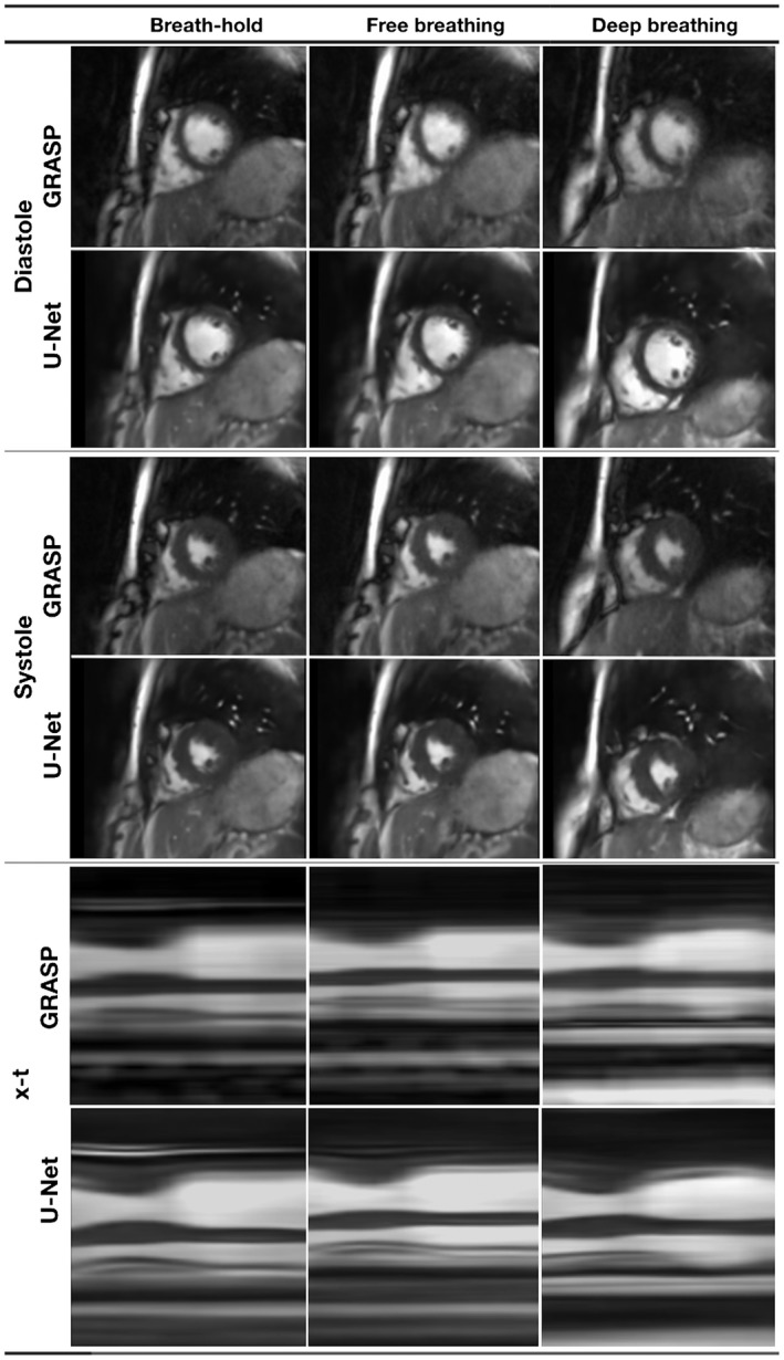
Figure 6
Full legend
Image quality in a single volunteer from the real‐time radial sequence reconstructed with GRASP and the residual U‐Net under different respiratory conditions (breath‐holding, free‐breathing and deep, rapid breathing) (see Supporting Information Video S4 for corresponding movie)
Show all 7 figures
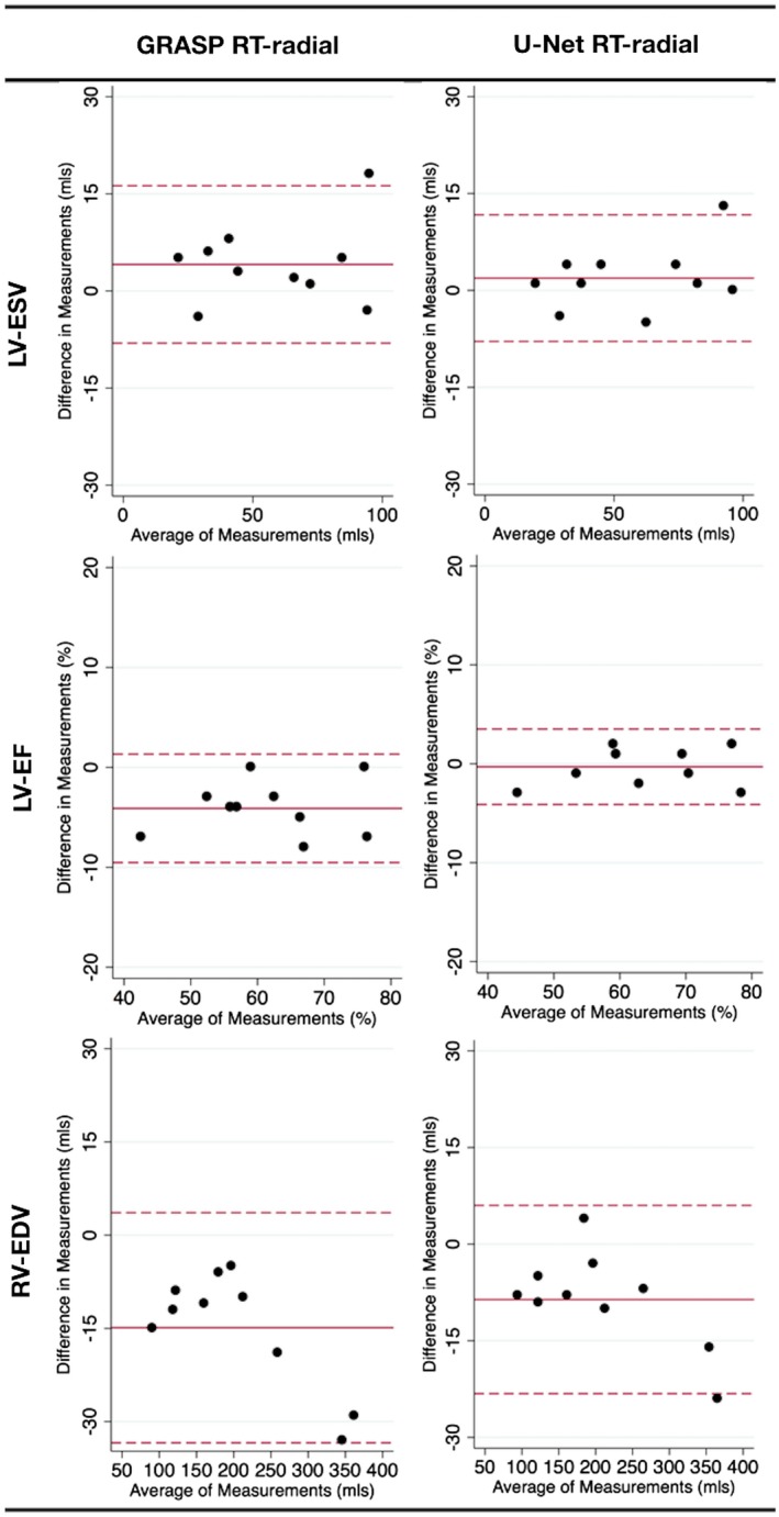
Figure 7
Full legend
Bland‐Altman plots showing ventricular volume results with significant biases. Comparison of ventricular volumes acquired with BH‐bSSFP to real‐time data reconstructed with GRASP and residual U‐Net. Solid red line shows mean difference, dashed lines shows ±2 SD
References30
- 1.
Grothues F, Smith GC, Moon J, et al. Comparison of interstudy reproducibility of cardiovascular magnetic resonance with two‐dimensional echocardiography in normal subjects and in patients with heart failure or left ventricular hypertrophy. Am J Cardiol. 2002;90:29‐34.
Not in our library10.1016/s0002-9149(02)02381-0 (opens in a new tab)
- 2.
Lustig M, Donoho D, Pauly JM. Sparse MRI: the application of compressed sensing for rapid MR imaging. Magn Reson Med. 2007;58:1182‐1195.
Not in our library10.1002/mrm.21391 (opens in a new tab)
- 3.
Kido T, Kido T, Nakamura M, et al. Assessment of left ventricular function and mass on free‐breathing compressed sensing real‐time cine imaging. Circ J. 2017;81:1463‐1468.
Not in our library10.1253/circj.cj-17-0123 (opens in a new tab)
- 4.
Haji‐Valizadeh H, Rahsepar AA, Collins JD, et al. Validation of highly accelerated real‐time cardiac cine MRI with radial k‐space sampling and compressed sensing in patients at 1.5T and 3T. Magn Reson Med. 2018;79:2745‐2751.
Not in our library10.1002/mrm.26918 (opens in a new tab)
- 5.
Wundrak S, Paul J, Ulrici J, Hell E, Rasche V. A small surrogate for the golden angle in time‐resolved radial MRI based on generalized Fibonacci sequences. IEEE Trans Med Imaging. 2015;34:1262‐1269.
Not in our library10.1109/tmi.2014.2382572 (opens in a new tab)
- 6.
Sandino CM, Dixit N, Cheng JY, Vasanawala SS. Deep convolutional neural networks for accelerated dynamic magnetic resonance imaging. In Proceedings of Medical Imaging meets Neural Information Processing Systems Conference, Long Beach, CA, 2017 p.19.
Not in our library
- 7.
Yang G, Yu S, Dong H, Slabaugh G, et al. DAGAN: deep de‐aliasing generative adversarial networks for fast compressed sensing MRI reconstruction. IEEE Trans Med Imaging. 2018;37:1310‐1321.
Not in our library10.1109/tmi.2017.2785879 (opens in a new tab)
- 8.
Qin C, Schlemper J, Caballero J, Price A, Hajnal JV, Rueckert D. Convolutional recurrent neural networks for dynamic MR image reconstruction. arXiv preprint arXiv 2017:171201751.
Not in our library10.1109/tmi.2018.2863670 (opens in a new tab)
- 9.
Ronneberger O, Fischer P, Brox T. U‐Net: convolutional networks for biomedical image segmentation. In Proceedings of the International Conference on Medical Image Computing and Computer‐Assisted Intervention, Munich, Germany, 2015 p. 234‐241.
Not in our library
- 10.
Jin KH, McCann MT, Froustey E, Unser M. Deep convolutional neural network for inverse problems in imaging. IEEE Trans Image Process. 2017;26:4509‐4522.
Not in our library10.1109/tip.2017.2713099 (opens in a new tab)
- 11.
Kang E, Min J, Ye JC. A deep convolutional neural network using directional wavelets for low‐dose X‐ray CT reconstruction. Med Phys. 2017;44:e360–e375.
Not in our library10.1002/mp.12344 (opens in a new tab)
- 12.
Han Y, Yoo J, Kim HH, Shin HJ, Sung K, Ye JC. Deep learning with domain adaptation for accelerated projection reconstruction MR. Magn Reson Med. 2018;80:1189‐1205.
Not in our library10.1002/mrm.27106 (opens in a new tab)
- 13.
Chan RW, Ramsay EA, Cheung EY, Plewes DB. The influence of radial undersampling schemes on compressed sensing reconstruction in breast MRI. Magn Reson Med. 2012;67:363‐377.
Not in our library10.1002/mrm.23008 (opens in a new tab)
- 14.
Abadi M, Barham P, Chen J, et al. TensorFlow: a system for large‐scale machine learning. In Proceedings of the 12th USENIX conference on Operating Systems Design and Implementation, Savannah, GA, 2016 p. 265‐283.
Not in our library
- 15.
Kingma DP, Ba J. Adam: a method for stochastic optimization. arXiv 2014:1412.6980.
Not in our library
- 16.
Larson P, Gurney PT, Nishimura DG. Anisotropic field‐of‐views in radial imaging. IEEE Trans Med Imaging. 2008;27:47‐57.
Not in our library10.1109/tmi.2007.902799 (opens in a new tab)
- 17.
Feng L, Grimm R, Block KT, et al. Golden‐angle radial sparse parallel MRI: combination of compressed sensing, parallel imaging, and golden‐angle radial sampling for fast and flexible dynamic volumetric MRI. Magn Reson Med. 2014;72:707‐717.
Not in our library10.1002/mrm.24980 (opens in a new tab)
- 18.
Tamir JI, Ong F, Cheng JY, Uecker M, Lustig M. Generalized magnetic resonance image reconstruction using the Berkeley advanced reconstruction toolbox. In Proceedings of ISMRM Workshop on Data Sampling and Image Reconstruction, Sedona, AZ, 2016 p.33.
Not in our library
- 19.
Afonso MV, Bioucas‐Dias JM, Figueiredo MA. An augmented Lagrangian approach to the constrained optimization formulation of imaging inverse problems. IEEE Trans Image Process. 2011;20:681‐695.
Not in our library10.1109/tip.2010.2076294 (opens in a new tab)
- 20.
McKenzie CA, Yeh EN, Ohliger MA, Price MD, Sodickson DK. Self‐calibrating parallel imaging with automatic coil sensitivity extraction. Magn Reson Med. 2002;47:529‐538.
Not in our library10.1002/mrm.10087 (opens in a new tab)
- 21.
Steeden JA, Atkinson D, Hansen MS, Taylor AM, Muthurangu V. Rapid flow assessment of congenital heart disease with high‐spatiotemporal‐resolution gated spiral phase‐contrast MR imaging. Radiology. 2011;260:79‐87.
Not in our library10.1148/radiol.11101844 (opens in a new tab)
- 22.
Rosset A, Spadola L, Ratib O. OsiriX: an open‐source software for navigating in multidimensional DICOM images. J Digit Imaging. 2004;17:205‐216.
Not in our library10.1007/s10278-004-1014-6 (opens in a new tab)
- 23.
Muthurangu V, Lurz P, Critchely J, Deanfield J, Taylor A, Hansen MS. Real‐time assessment of right and left ventricular volumes and function in patients with congenital heart disease by using high spatiotemporal resolution radial k‐t SENSE. Radiology. 2008;248:782‐791.
Not in our library10.1148/radiol.2482071717 (opens in a new tab)
- 24.
Schlemper J, Caballero J, Hajnal JV, Price A, Rueckert D. A deep cascade of convolutional neural networks for MR image reconstruction In: Niethammer M, Styner M, Aylward S, Zhu H, Oguz I, Yap PT, Shen D, editors. Information processing in medical imaging: 25th International Conference, IPMI 2017, Boone, NC, USA, June 25–30, 2017, Proceedings. Cham: Springer International Publishing; 2017 p. 647‐658.
Not in our library
- 25.
Hammernik K, Klatzer T, Kobler E, et al. Learning a variational network for reconstruction of accelerated MRI data. Magn Reson Med. 2018;79:3055‐3071.
Not in our library10.1002/mrm.26977 (opens in a new tab)
- 26.
Yu S, Dong H, Yang G, Slabaugh G, et al. Deep de‐aliasing for fast compressive sensing MRI. arXiv preprint arXiv 2017:170507137.
Not in our library
- 27.
Zhao H, Gallo O, Frosio I, Kautz J. Loss functions for image restoration with neural networks. IEEE Trans Comput Imaging. 2017;3:47‐57.
Not in our library
- 28.
Lim B, Son S, Kim H, Nah S, Lee KM. Enhanced deep residual networks for single image super‐resolution. In Proceedings of the IEEE Conference on Computer Vision and Pattern Recognition (CVPR) Workshops, Honolulu, HI, 2017 p. 3.
Not in our library
- 29.
Mardani M, Gong E, Cheng JY, et al. Deep generative adversarial networks for compressed sensing automates MRI. arXiv preprint arXiv. 2017:170600051.
Not in our library
- 30.
Hammernik K, Knoll F, Sodickson DK, Pock T L2 or not L2: impact of loss function design for deep learning MRI reconstruction. In Proceedings of the 25th Annual Meeting of ISMRM, Honolulu, HI, 2017. Abstract 0687.
Not in our library
Reads
Read by people on this page
None yet.
Checked or read by AI through naulon
None yet.
A person counts once a day per paper. We keep no cookie and no address: a reader is a hash that changes every day. Any month under five reads shows no number.
Are you an author?
Is this your paper?
Claim it to add your note and be paid when agents cite it. Free.